
|
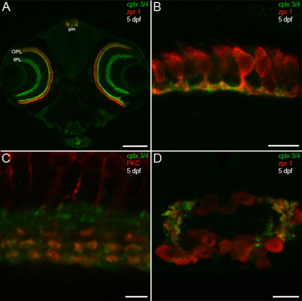
Fig. 3 Predilection of complexin 3/4 for ribbon presynaptic terminals in the larval zebrafish visual system. (A) A confocal projection of a 5-dpf zebrafish transverse section stained with the pan-complexin 3/4 antibody (green) and the zpr 1/FRet 43 antibody (red), which labels double cone photoreceptors, reveals complexin 3/4 immunoreactivity throughout the retinal plexiform layers (OPL, IPL) and in the pineal organ (pin). Complexin 3/4 is absent from the retinal nuclear layers and the medial region of the pineal organ, which contains photoreceptor somata. (B) A high-magnification confocal projection of double cone photoreceptors (red) in the retinal OPL labeled with the complexin 3/4 antibody (green) shows that some of the complexin 3/4 immunoreactivity in the larval zebrafish OPL is found in double cone terminals. (C) Double-labeling with anti-protein kinase C (red) reveals overlap of complexin 3/4 (green) in ON bipolar cell terminals in the retinal IPL. (D) A confocal projection of a transverse section through the pineal organ reveals zpr 1/FRet 43-positive photoreceptors (red) and complexin 3/4 (green) in processes and terminals at the lateral border. Sections incubated with secondary antibodies alone exhibit background immunofluorescence in the retina and pineal (data not shown). Scale bars: 125 μm (A); 10 μm (B, D); 5 μm (C).