
Image
|
Figure Caption
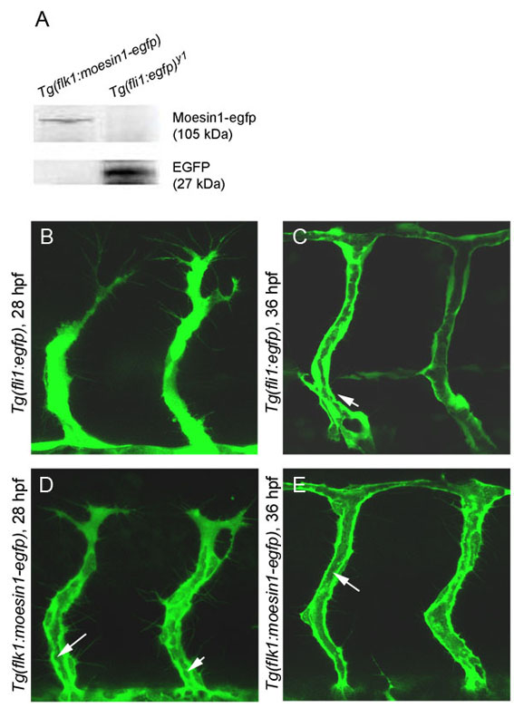
Fig. S2 Comparison of the ability to detect the primary lumen between Tg(fli1:egfp)y1 embryos and Tg(flk1:moesin1-egfp) embryos. (A) Western blots were performed as described (Link et al., 2006) using 30 hpf embryos. Western blot analysis of the Moesin1-EGFP fusion protein. (B,D) At 28 hpf, vacuoles contributing to the formation of the primary lumen (arrow) are clearly visible in the Tg(flk1:moesin1-egfp) but not the Tg(fli1:egfp)y1 line. (C,E) At 36 hpf, the primary lumen in the ISVs (arrows) is observed in both lines.
Acknowledgments
This image is the copyrighted work of the attributed author or publisher, and
ZFIN has permission only to display this image to its users.
Additional permissions should be obtained from the applicable author or publisher of the image.
Full text @ Development