
|
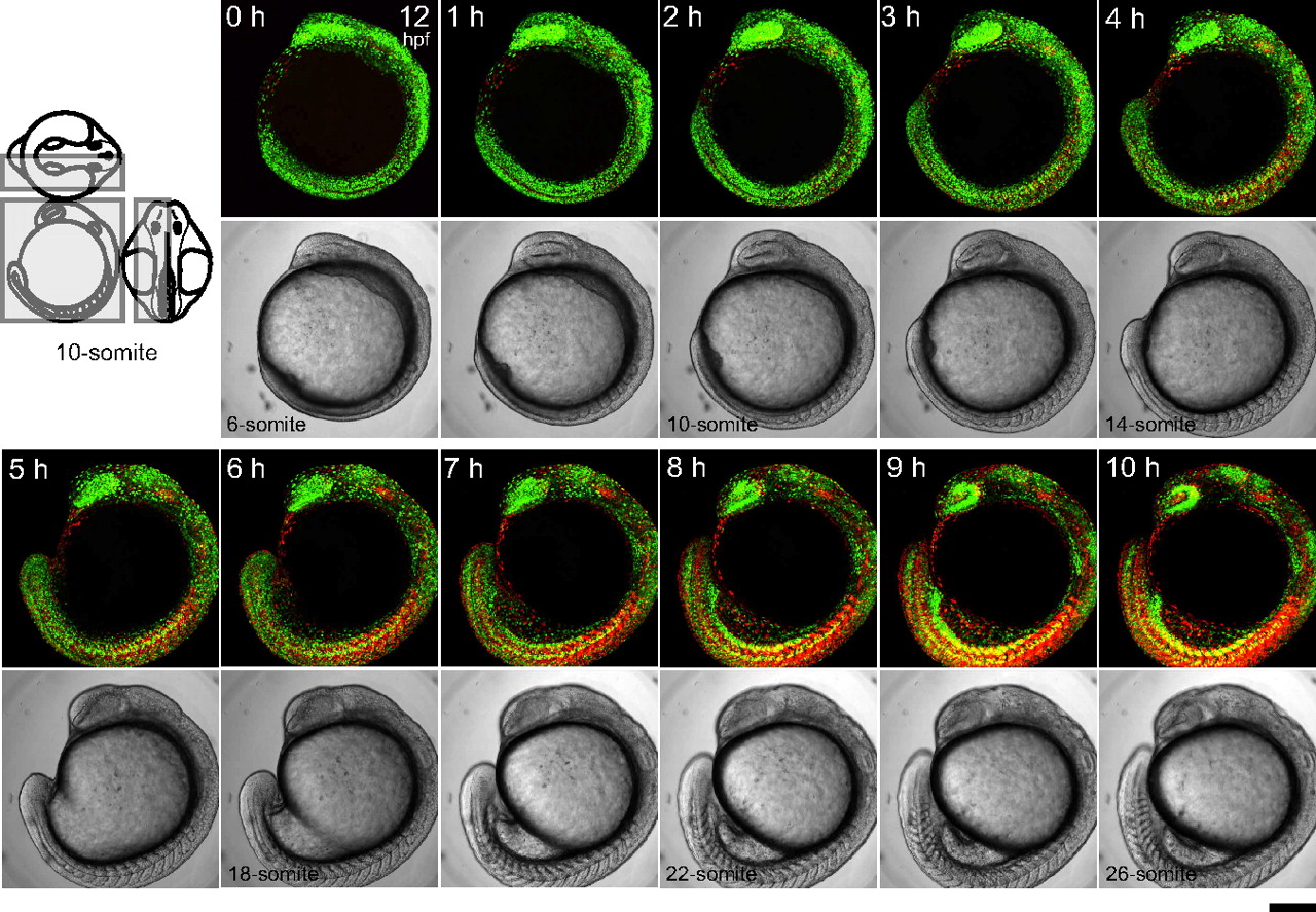
Fig. 2 Time-lapse imaging of a Cecyil embryo during segmentation. Three-dimensional time-lapse imaging was performed to collect fluorescence images from the left half of the embryo at 10-min intervals using an Olympus FV1000 upright confocal microscope equipped with an objective lens (x10 N.A. 0.3). A dechorionated embryo in the early segmentation period at 12 hpf (6-somite stage) was mounted with the left side up in a chamber containing 0.3% agar. Since the sample was kept at less than 28 °C during observation, developmental stages cannot be accurately expressed in hpf. Due to z-stacking, green and orange signals at different z-positions merge to generate yellow signal. Note that zFucci does not yield yellow fluorescence at the G1/S transition, whereas the original Fucci in mammalian cells does. The scanned region is indicated by the gray box on the three views of an embryo at the 10-somite stage. (Scale bar, 200 μm.)