
|
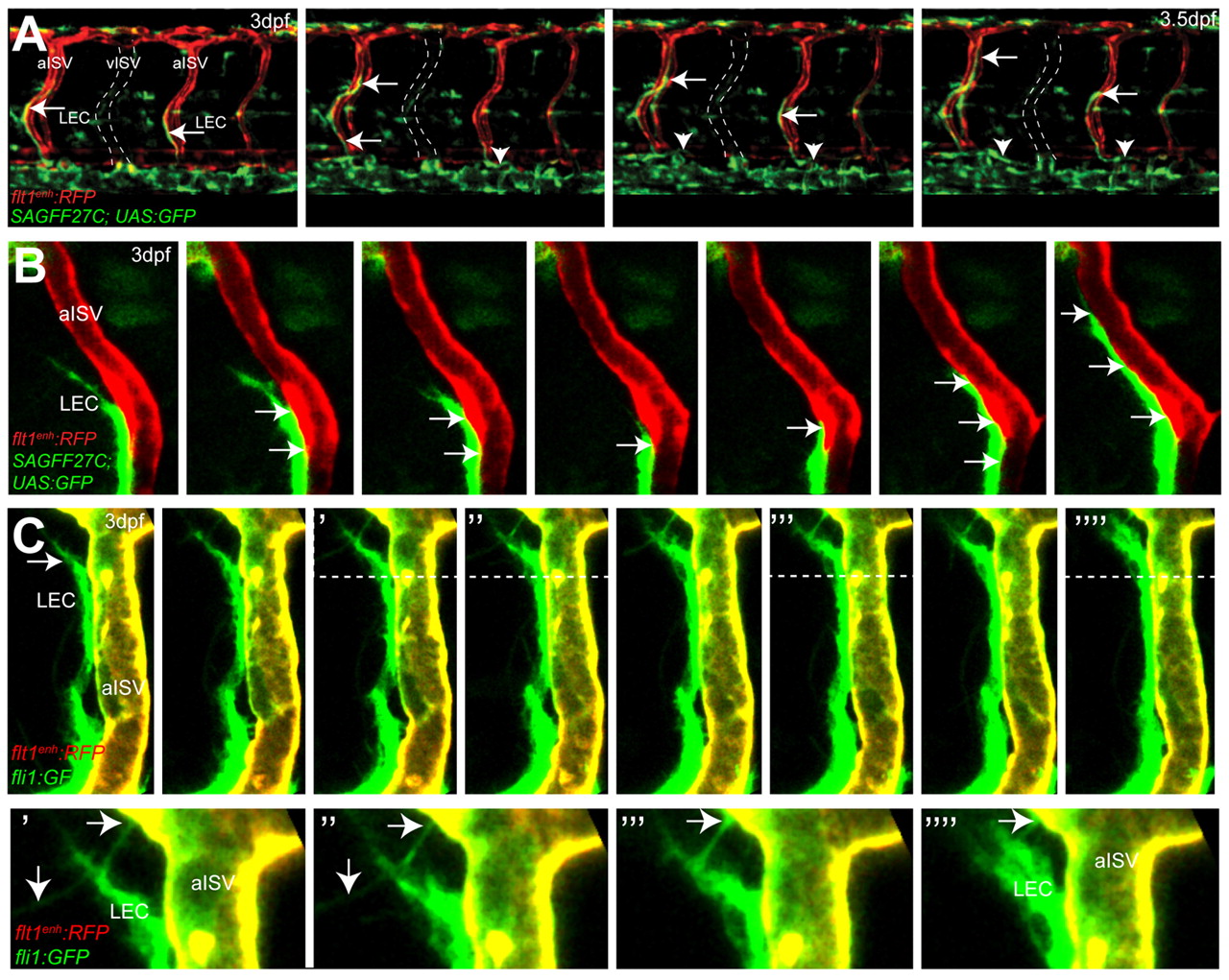
Fig. 3 Migration of lymphatic precursors along intersegmental arteries. (A) Still images of flt1enh:RFP;SAGFF27C;UAS:GFP triple transgenic embryos reveal migration of SAGFF27C;UAS:GFP+ LECs (arrows) along flt1enh:RFP+ arteries and establishing the TD (arrowheads) in the trunk. The position of a vISV is indicated by broken lines. (B) Still images of migrating SAGFF27C;UAS:GFP+ LECs suggest direct contact (arrows) between the aISV and the LEC. (C) Still images of migrating fli1a:GFP+ LECs and filopodia formation (indicated by arrows) at the leading edge along the surface of a flt1enh:RFP+; fli1a:GFP+ artery at 3 dpf. Areas above the broken lines are shown at higher magnification in the bottom row.