
|
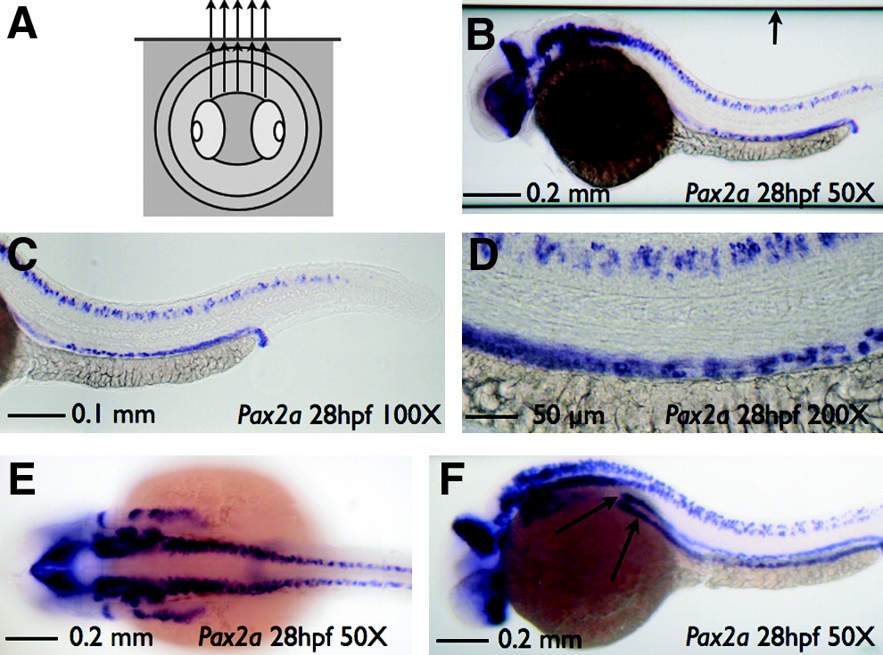
Fig. 3 SCORE imaging of a fixed specimen. Imaging of a pax2a whole-mount in situ hybridization embryo and capillary housing in 75% glycerol. (A) Cartoon depicting the use of glycerol both inside and outside of the glass tube producing an undistorted image of a fixed embryo. (B) Sagittal image of embryo with pax2 staining at 50x magnification shows no distortion (also see Supplemental Movie S4, available online at www.liebertonline.com). Note the edges of the capillary (arrow) that can be readily cropped for publication presentation. Sagittal embryo image of pax2 staining at 100x (C) or 200x (D) magnification shows no distortion. (E) Coronal image of embryo at 50x magnification. (F) Angled image of pax2 staining at 50x magnification. Rotation is angled slightly (∼30°) to show a more detailed view of kidney tubule staining (arrows).