
|
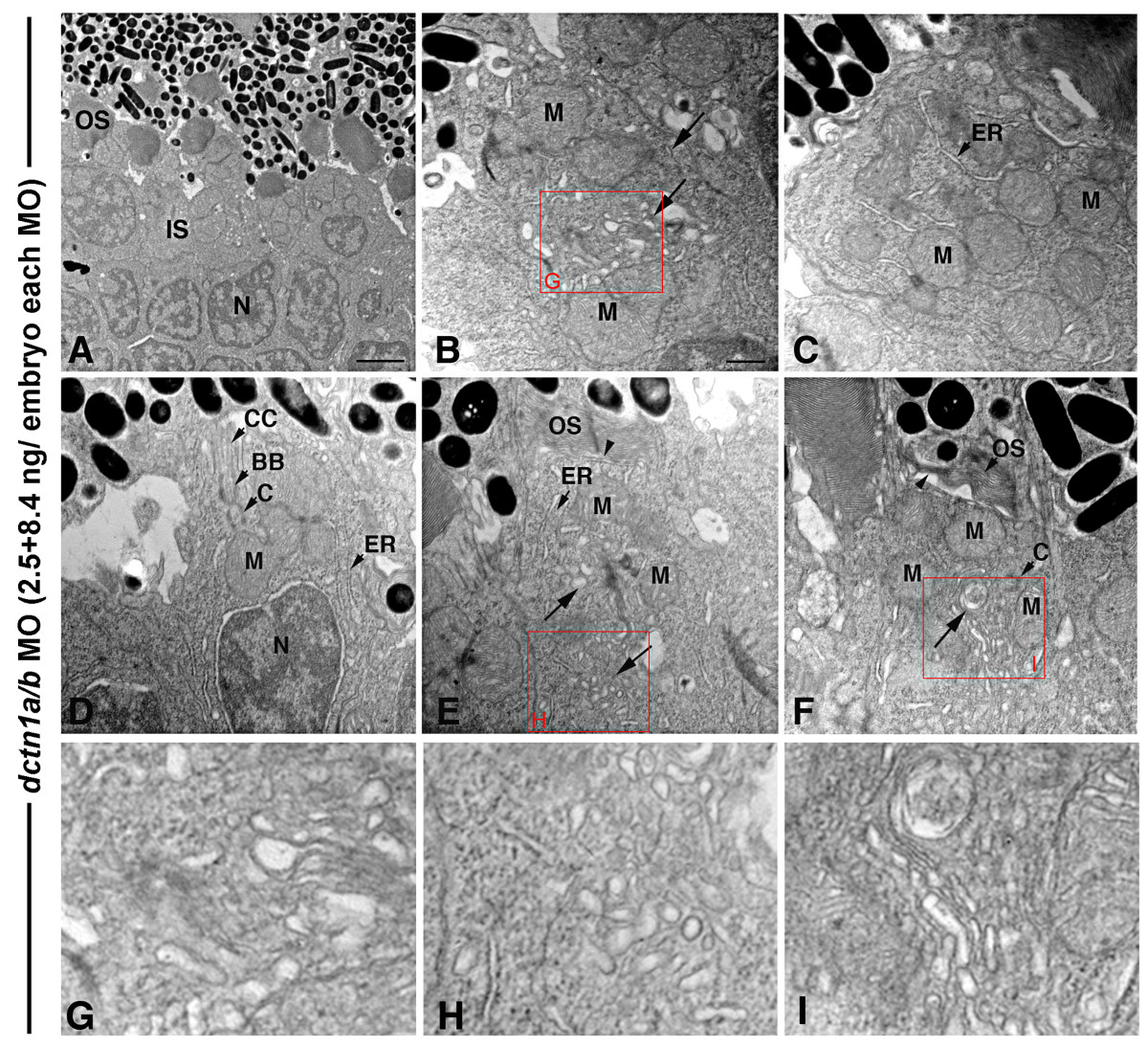
Fig. 12 Double dnct1a/b knock-down primarily affects the inner segment. (A) TEM view of the central retina of a dctn1a/b morphant at 3 days showing normal, albeit short, outer segment structures. (B-F) TEM views of individual photoreceptors at the peripheral dctn1a/b morphant retina with inner segment polarization defects, including a dispersion of mitochondria (M) within that region (B, C, F), lack of a distinct myoid region (D) and positioning of the ER/Golgi complex among dispersed mitochondria (arrows in (B, E, F)). In addition, accumulation of Golgi associated vesicles in the inner segment (arrow in (B, E, F)) and shortened outer segment structures (arrowheads) were also seen (E, F). (G-I) Higher magnification insets of the red boxed regions. For morpholino (MO) injections, dnct1a splice MO was used at 2.5 ng/embryo, while the dnc1b splice MO was used at 8.4 ng/embryo (for a total MO concentration of 10.9 ng/embryo). BB, basal body; C, centriole; CC, connecting cilium; ER; endoplasmic reticulum; IS, inner segment; M, mitochondria; OS, outer segment. Scale bar: 2 μm in (A); 0.5 μm in (B-F).