
|
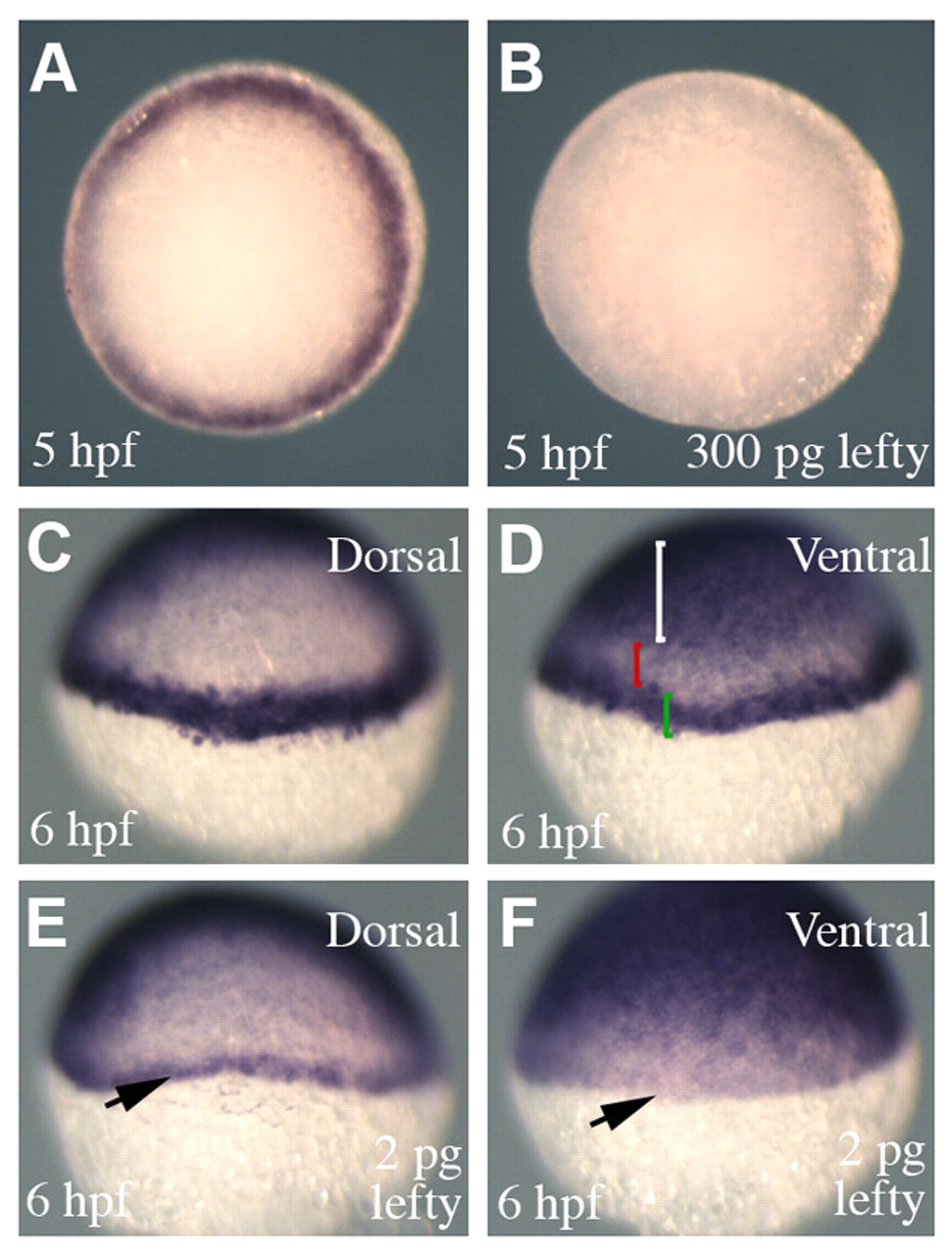
Fig. 6 Attenuation of nodal signalling in Tg(-1 kb ntl:CFP) embryos. (A,B) Animal pole views showing transgene expression in Tg(-1 kb ntl:CFP) embryos. Injection of high concentrations of lefty (300 pg) completely abolished transgene expression (B) (100%, n=68). (C,D) A transgenic embryo processed to show transgene and gata2 expression, which marks the ventral ectoderm. (D) The white bracket highlights gata2 expression in the ventral ectoderm and the green bracket shows transgene expression in the margin. The red bracket shows a region that separates the expression domains of gata2 and the transgene. (E,F) A transgenic embryo injected with a low concentration of lefty (2 pg). (E) On the dorsal side of the embryo the transgene is expressed in fewer cell tiers compared with uninjected embryos (C). However, within the same embryo, transgene expression is abolished in the ventral margin (arrow) (expression pattern observed in 43%; in 50% transgene expression was completely abolished, n=60).