
|
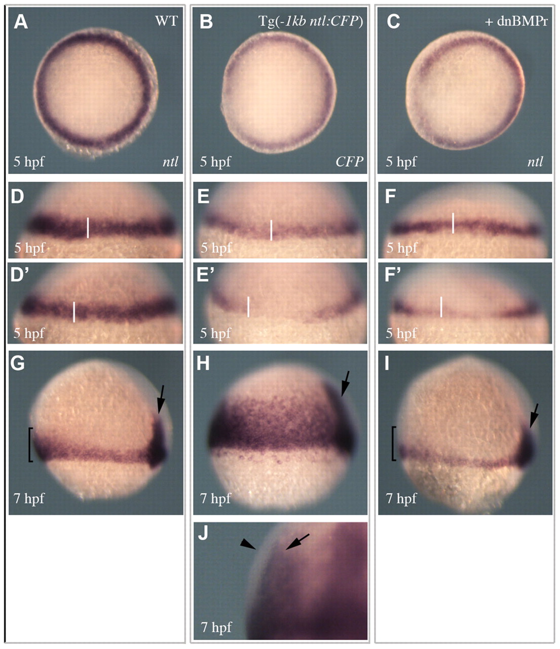
Fig. 5 Spatial and temporal regulation of ntl expression. (A,D,D′,G) ntl expression in wild-type embryos. (B,E,E′,H,J) Expression of the -1 kb ntl promoter transgene. (C,F,F′,I) The expression of ntl in embryos injected with a dnBMPr. (A-C) Animal pole views of expression at 5 hpf. (D-F′) Views of expression on opposing sides of individual embryos at 5 hpf (transgene expression 84%, n=51 and dnBMPRr injected 69%, n=46). (G-I) Lateral views of expression at 7 hpf. Arrows indicate expression in the developing notochord. Brackets in G and I highlight the enriched expression in the ventral margin of wild-type embryos, which is not observed in embryos injected with the dnBMPr (92%, n=37). (J) A magnified lateral view of a 7 hpf embryo shows the transgene is expressed in the underlying endodermal cells (arrow) and not the outer mesodermal cells (arrowhead) (100%, n=52).