
|
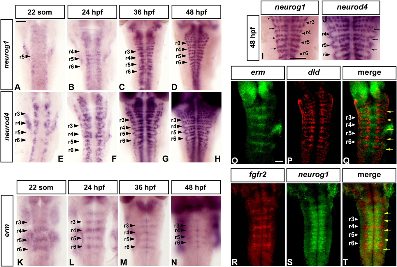
Fig. 1
FGF Signaling Is Restricted to Nonneurogenic Regions in the Zebrafish Hindbrain
Dorsal views of flat mounted embryos, anterior to the top, at the indicated stages. Following in situ hybridization, embryos of the same batch were developed for the same amount of time. Arrowheads indicate segment centers; arrows point at hindbrain boundaries. Scale bars, 50 μm. (A–H) Time course of neurog1 (A–D) and neurod4 (E–H) expression from 22 somites to 48 hr. (I and J) Higher-power views showing the spatial restriction of neurogenesis marked by neurog1 (same embryo as [D]) and neurod4 ([J], same embryo as [H]). (K–N) erm expression. (O–T) Double fluorescent in situ hybridization using probes for erm and dld (O–Q), and fgfr2 and neurog1 (R–T). Images shown are a merge of confocal stacks through the hindbrain at 36 hpf.
Reprinted from Developmental Cell, 18(1), Gonzalez-Quevedo, R., Lee, Y., Poss, K.D., and Wilkinson, D.G., Neuronal Regulation of the Spatial Patterning of Neurogenesis, 136-147, Copyright (2010) with permission from Elsevier. Full text @ Dev. Cell