
|
Fig. 5
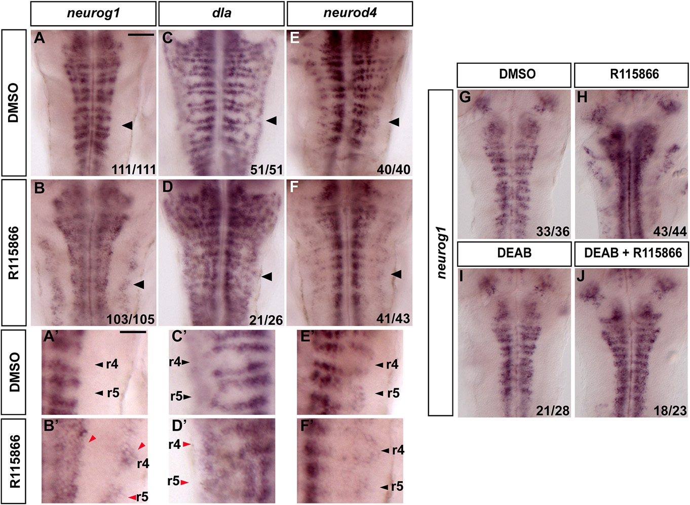
Blocking Cyp26 Activity Results in Premature Neurogenesis
(A–F) In situ hybridization of 40 hpf embryos to detect expression of neurog1 (A and B), dla (C and D) or neurod4 (E and F) in DMSO- or R115866-treated embryos. Treatments were started at 24–26 hr. Black arrowhead points at r5. Scale bar, 50 μm. (A′–F′) Higher-power views of r4 and r5 shown in A–F (black arrowheads).
Red arrowheads indicate ectopic proneural expression. Scale bar, 25 μm.
(G–J) Blocking RA signaling with DEAB partially rescues loss of Cyp26. In situ hybridization of 36 hpf embryos to detect expression of neurog1 in DMSO (G), R115866 (H), DEAB (I), or R115866 + DEAB (J) -treated embryos.
Reprinted from Developmental Cell, 18(1), Gonzalez-Quevedo, R., Lee, Y., Poss, K.D., and Wilkinson, D.G., Neuronal Regulation of the Spatial Patterning of Neurogenesis, 136-147, Copyright (2010) with permission from Elsevier. Full text @ Dev. Cell