
|
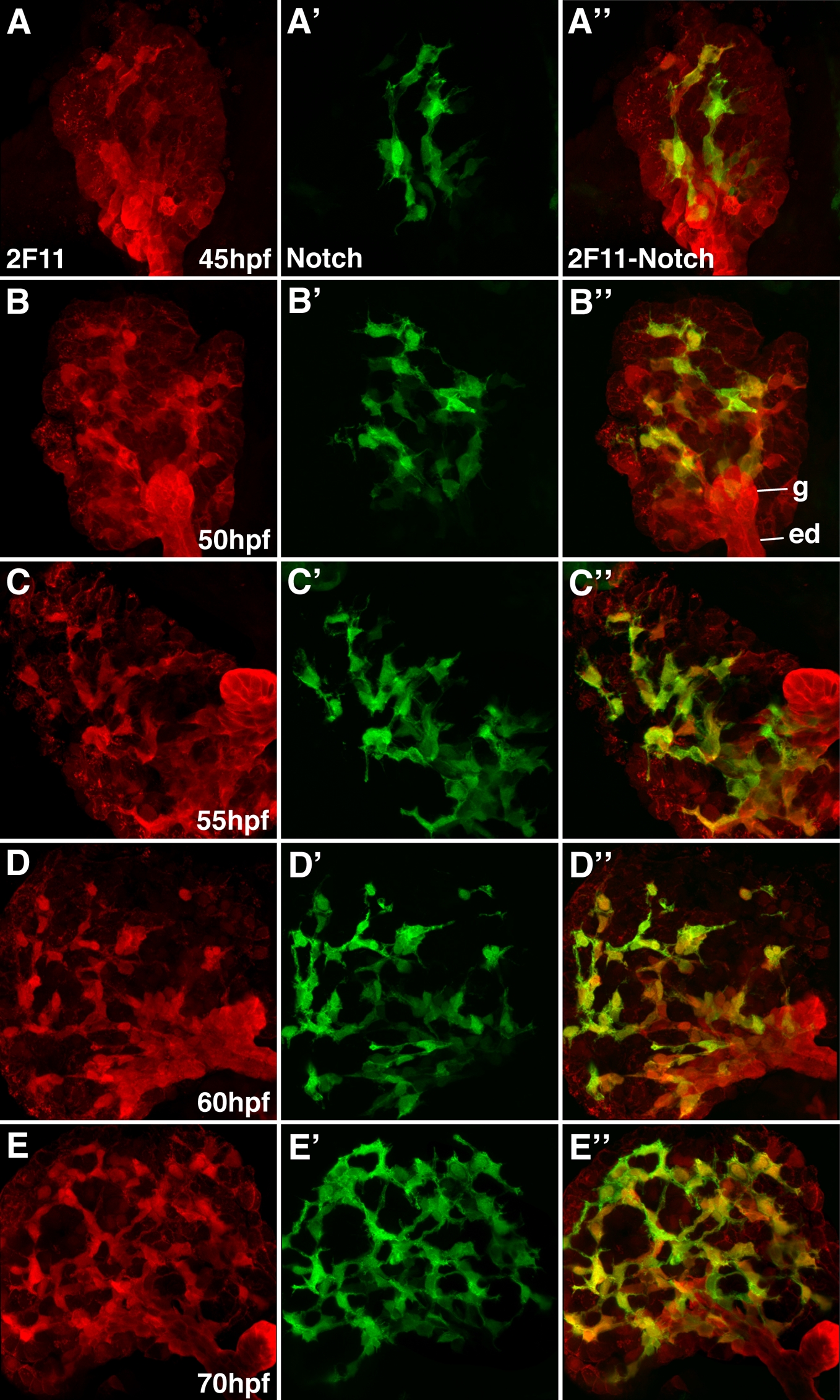
Fig. S5 Notch reporter expression in developing intrahepatic biliary cells: Whole mount confocal images (63x) through the liver of developing Notch reporter larvae stained with the 2F11 antibody (A-D) and a GFP antibody (A′-D′) with overlap of the two markers (A″-D″). GFP-positive biliary epithelia are first detected at 45 hpf (A′). At this stage, scattered 2F11-positive cells are present in the liver with only partial overlap with GPF. From 50-70 hpf, there is progressive increase in the number of GFP-positive cells. AT 50 hpf, there is significant overlap between the GFP and 2F11 epitope in the biliary cells. There is nearly complete overlap between these markers at 60 and 70 hpf. The gallbladder (g) and extrahepatic duct (ed) remain GPF negative.