
|
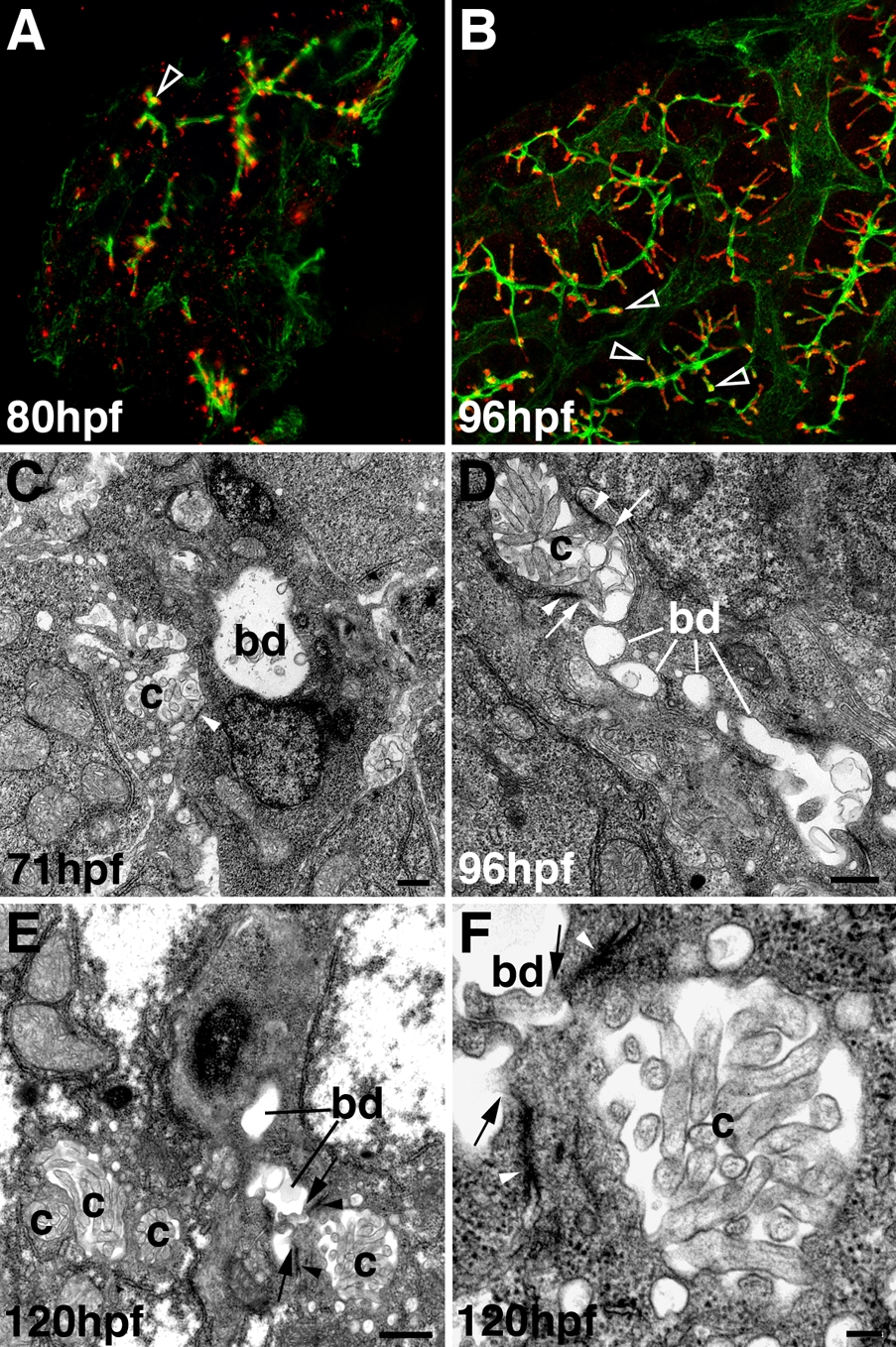
Fig. S2 Development of hepatocyte canaliculi and intrahepatic biliary network. A, B: Confocal projections through the liver of an 80-hpf (A) and 96-hpf (B) larva immunostained with Keratin-18 (green) and Mdr (red) antibodies. Compared with double immunostains of 5-dpf larvae (Fig. 2F″), there is much less overlap of the Mdr-1 and Keratin-18 epitopes at these developmental stages (open arrowheads). C-F: Transmission electron micrographs showing the canalicular-terminal duct junction (arrows) in 71-hpf (C), 96-hpf (D), and 120-hpf (E, F) larvae. bd, bile duct; c, canaliculus; arrowhead, tight junctions. Bar = 500 nm in C-E, 100 nm in F