
|
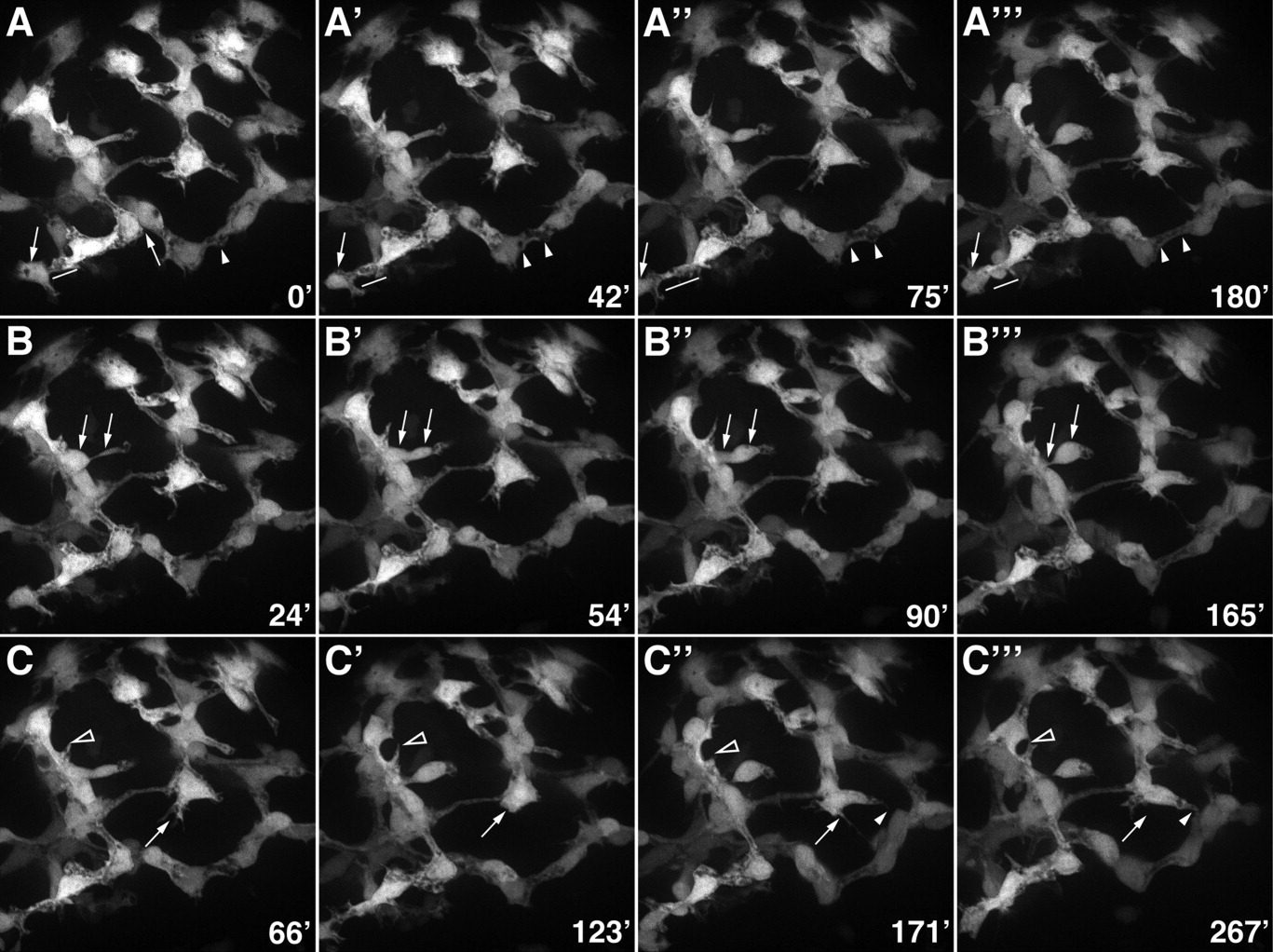
Fig. 8 Morphogenesis of the intrahepatic biliary network. Confocal projections through the liver that was dissected from a 76-hpf Notch reporter larva and cultured for 4.5 hr. Numbers in the bottom right corner indicate duration of culture in minutes. A: These images show progressive lengthening and then shortening of the cytoplasmic bridge between two adjacent biliary cells (arrows). Fusion of cytoplasmic vesicles leading to the formation of a ductal lumen within two adjacent cells is also evident (arrowheads). B: These images show the migration of a single biliary cell via a cytoplasmic extension (arrows). C: These images show the dynamic pattern of filopodia extension and retraction from two biliary cells (arrows and arrowheads).