
|
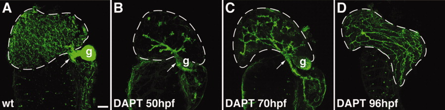
Fig. 3 Notch signaling is required for expansion of the intrahepatic biliary ductal network. A-D: Confocal projections through the liver of a control 120-hpf larva and 120-hpf larvae treated with DAPT beginning at 50 hpf (B), 70 hpf (C), and 96 hpf (D) that were stained with the keratin-18 antibody. These images show that at all stages examined, expansion of the biliary network is disrupted when Notch signaling is inhibited by DAPT. Development of the extrahepatic duct (arrows) and gallbladder (g) are not affected by Notch inhibition. These structures are out of the plane of focus in the 96-hpf larva.