
|
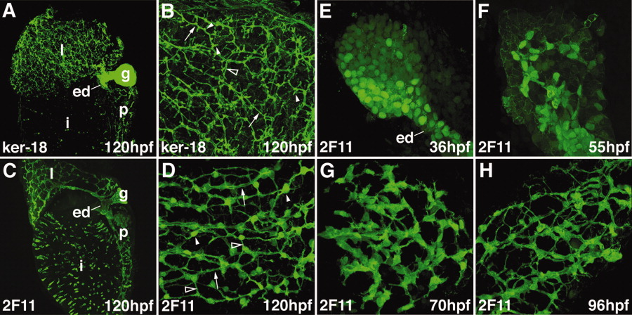
Fig. 1 Zebrafish intrahepatic biliary development. A, B: Confocal projections through the liver of a 120-hpf larvae stained with the anti-Keratin 18 antibody. The low-power image (A) shows the intrahepatic biliary network (l), extraehepatic ducts (ed), and gallbladder (g). The high-power image (B) shows that the keratin-18 protein detects 3 classes of intrahepatic ducts: long ducts (arrow), interconnecting ducts (open arrowhead), and terminal ductules (arrowhead). C, D: Confocal projections through the liver of a 120-hpf larvae stained with the 2F11 antibody. The low-power image (C) shows that the 2F11 epitope is presented within the intrahepatic (l) and extrahepatic (ed) ductal systems as well as the gallbladder (g). The high-power image (D) shows that the 2F11 epitope is present in the nucleus of the biliary epithelial cell (arrowhead). This epitope is also detected on the long ducts (arrow) and interconnecting ducts (open arrowhead) but not the terminal ductules. E-H: Developmental pattern of the 2F11 epitope in the intrahepatic biliary system between 36 and 96 hpf.