
|
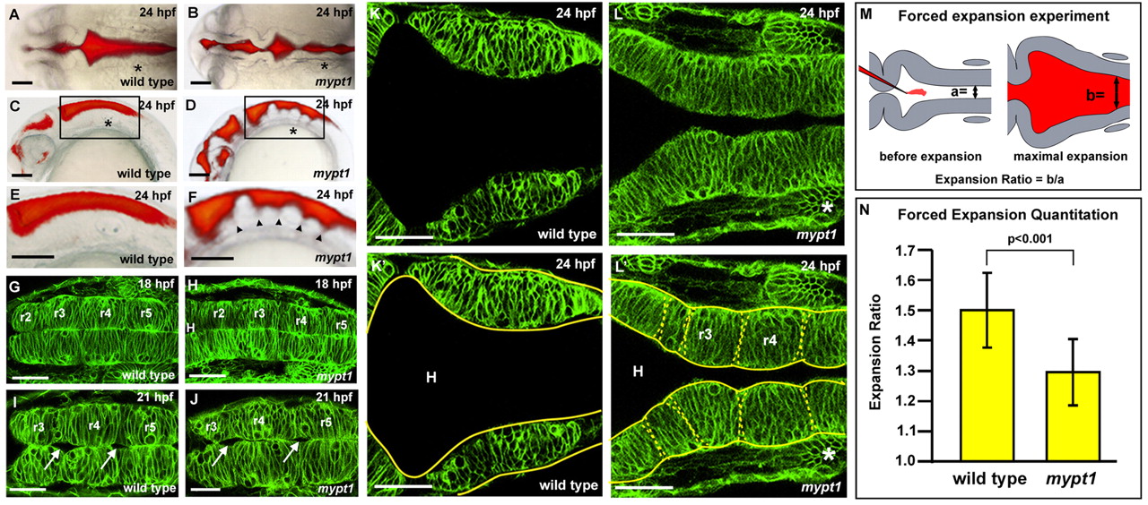
Fig. 2 The mypt1 mutant phenotype. (A-F) Wild-type (A,C,E) and mypt1 mutant (B,D,F) zebrafish brain ventricle injections at 24 hpf. Views are dorsal (A,B) or lateral (C,D). (E,F) Higher magnification of boxed regions from C and D, respectively. Arrowheads indicate rhombomere boundaries. (G-L′) Live confocal imaging of the hindbrain in wild type (G,I,K,K′) and mypt1 mutants (H,J,L,L′) after memGFP mRNA injection. (G,H) Wild type (G) and mypt1 mutants (H) at 18 hpf. (I,J) Wild type (I) and mypt1 mutants (J) at 21 hpf showing similar morphology and hindbrain openings (arrows). (K-L′) Wild type (K) and mypt1 mutants (L) at 24 hpf. (K′,L′) The same images as in K and L but with neuroepithelial tissue outlined in yellow. Dashed lines denote rhombomere morphology (L′). n>10 for all panels. mypt1 mutants have a normal neural tube, suggesting maternal gene expression is sufficient for brain development until the end of neurulation. Consistent with this, inhibition of Mypt1 expression using a start site MO led to a disorganized neural tube (data not shown), whereas a splice site MO (see Fig. S1 in the supplementary material) showed a phenotype only after the neural tube had formed. (M) Schematic of the hindbrain before and at maximum forced ventricle expansion by high-pressure injection of fluid; a and b indicate the regions that were measured. (N) Quantitation of the expandability of the hindbrain neuroepithelium in wild type and mypt1 mutants. Two independent experiments were averaged; wild type, n=12; mypt1, n=15. Data are represented as an expansion ratio ± s.d. Results are significantly different (P<0.001, Mann-Whitney U-test). Asterisks denote the ear. Anterior is to the left. H, hindbrain ventricle. Scale bars: 100 μm in A-F; 50 μm in G-L′.