
|
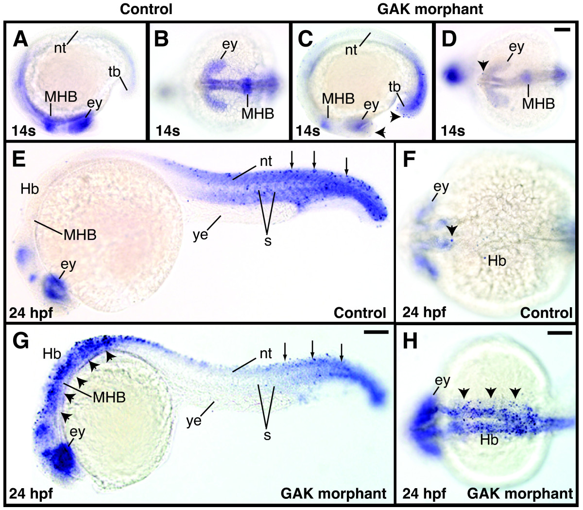
Fig. 8 Programmed cell death in wild-type and GAK morphant embryos during development. (A, C) Lateral and (B, D) dorsal views of TUNEL-stained (A, B) wild-type and (C, D) GAK morphant embryos at the 14-somite stage. At this stage, the control embryos display no detectable apoptosis, whereas GAK morphants have a low level of programmed cell death (indicated by black arrowheads). (E, G) Lateral and (F, H) dorsal views of TUNEL-stained (E, F) wild-type and (G, H) GAK morphants at 24 hpf. While the control animal exhibits a low level of apoptosis in anterior brain and the posterior body regions (arrowheads), GAK morphants exhibit a high level of apoptotic cell death in the brain, as well as in the neural tube (arrowheads). However, as compared to the control wild-type embryo, no obvious increase in apoptosis was observed in the posterior region of GAK morphants (indicated by arrows). In all the lateral views, anterior is to the left and dorsal is up. ey, eye; Hb, hindbrain; MHB, mid-hindbrain boundary; nt, neural tube; s, somites; tb, tailbud. Scale Bar, 100 m.