
|
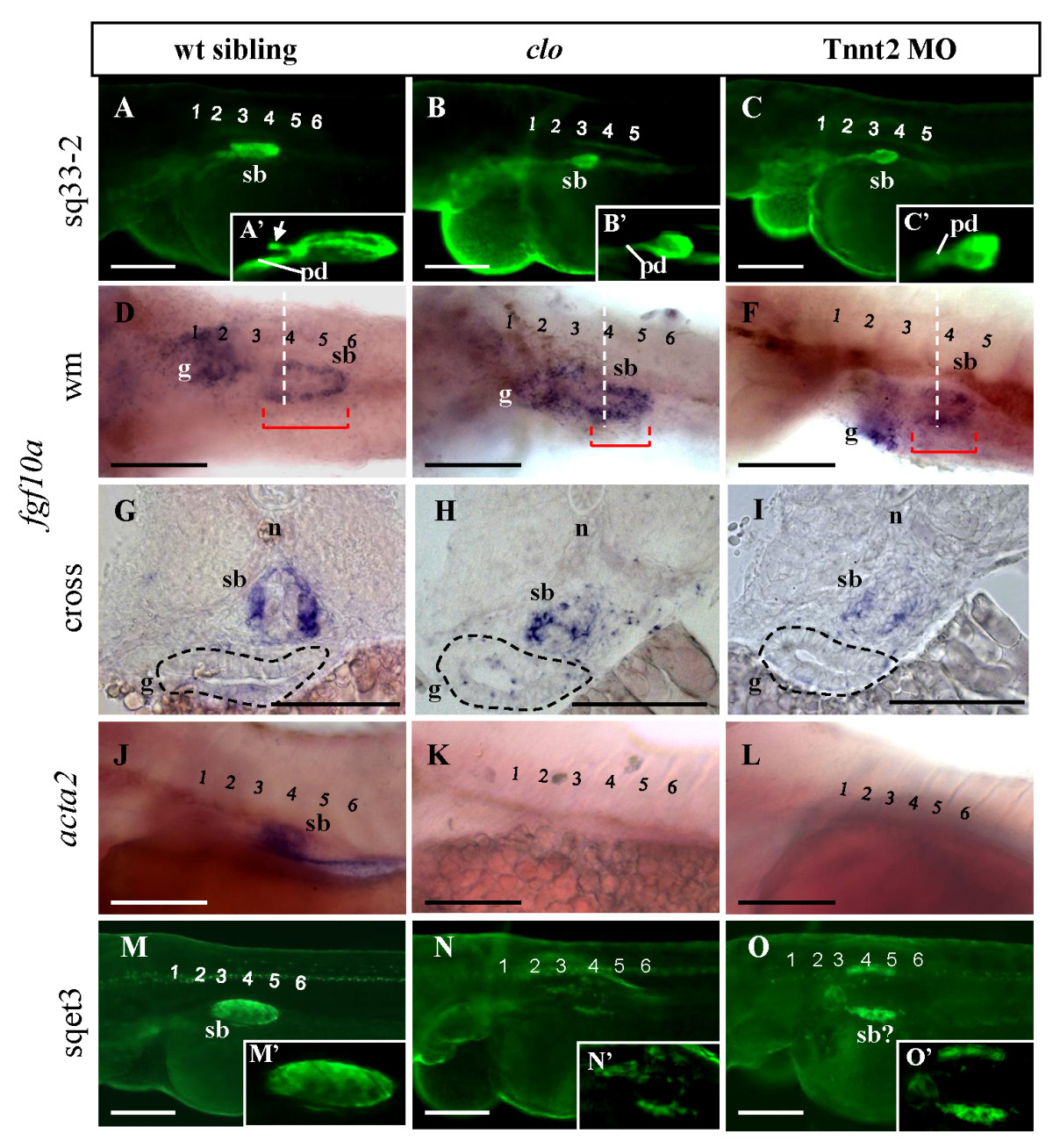
Fig. 3 The role of ECs and blood circulation on swimbladder growth. (A - C) EGFP expression in control, clo-/-, mutant, and Tnnt2 morphants on the background of Et(krt4:EGFP)sq33-2 at 72 hpf. Swimbladder appeared smaller in larvae without endocardium and endothelium (clo-/-, B) and without cardiac function (Tnnt2 MO, C) as compared to wild type (A). (A′ - C′) 2.5x magnification of the swimbladder in (A - C) respectively. The white arrow indicates the primordium of swimbladder anterior chamber. (D - F) Presence of swimbladder mesenchyme at 72 hpf as detected by fgf10a expression in wild type, clo-/- mutant, and Tnnt2 morphant. (G - I) Cross section at the levels indicated by white dashed lines in panel (D - F) to show mesenchymal fgf10a expression in wild type, clo-/- mutant, and Tnnt2 morphant larvae at 72 hpf. Red bar indicates swimbladder length, Gut (g) is demarcated by black dashed line. (J - L) Analysis of acta2 expression in swimbladder in wild type (J), clo-/- mutant (K) and Tnnt2 morphant (L) at 72 hpf. (M - O) EGFP expression in swimbladder outer mesothelium in wild type (M), clo-/- mutant (N), and Tnnt2 morphant (O) at 72 hpf. Note the severe reduction of outer mesothelium in clo-/- mutant and its disorganization into two lateral stripes in Tnnt2 morphant. (M′ - O′) 2.5x magnification of the swimbladder in (M - O) respectively. Numbers indicate anterior somites. Abbreviations: pd, pneumatic duct; n, notochord; sb, swimbladder. Scale bars: 250 μm.