
|
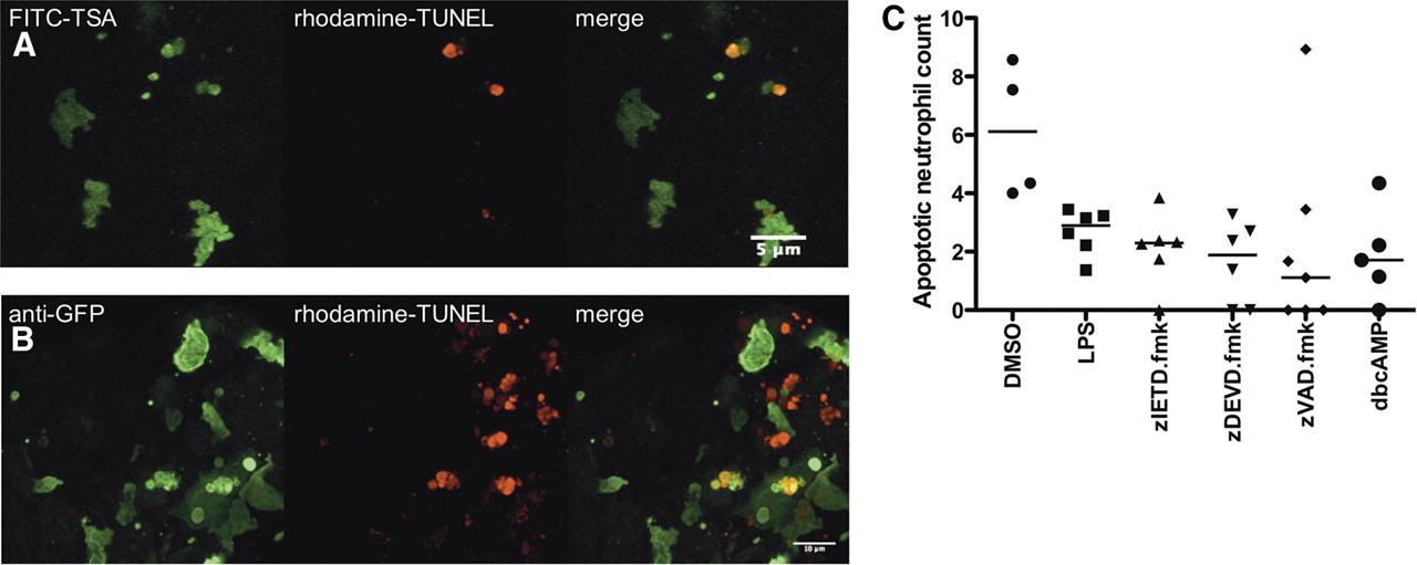
Fig. 4 Dual staining for neutrophil-specific mpx and DNA nick-ends (TUNEL) provides biochemical confirmation of neutrophil apoptosis during resolution of neutrophilic inflammation in unmanipulated zebrafish embryos. Seven dpf zebrafish were injured and fixed 12 h later in 4% paraformaldehyde at 4°C. Neutrophils were revealed by (A) FITC-TSA staining (wild-type fish) for peroxidase activity or (B) immunostaining for GFP (mpx:GFP fish), in combination with TUNEL staining with the ApopTag Red kit. Dual-positive cells representing apoptotic neutrophils can be seen in the right panel of A and B. The images shown are maximum-intensity projected images of at least 20 sections in the Z dimension. Images were taken using a x60 Plan Apo Oil immersion NA1.40 (Olympus). Colocalization of fluorescence in single sections can be seen in Supplemental Movie 3. (C) Larvae at 10 dpf were injured as described previously and treated with the compounds indicated. At 12 h after injury, larvae were fixed and stained for colocalization of neutrophil (FITC-TSA) and apoptotic (TUNEL) markers as above. Dual-positive cells and total neutrophil count were scored from confocal images. Data shown are percent neutrophils costaining with TUNEL markers (i.e., apoptotic neutrophils). P < 0.05 for all treatments compared with DMSO control by one-way ANOVA with Dunnett’s post-test correction. zVAD.fmk, zVD.fmk.