
|
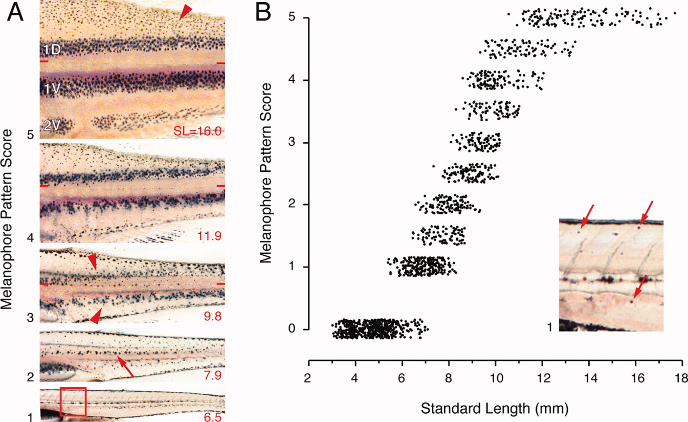
Fig. 25 Body melanophore pattern. A: Pigment pattern metamorphosis in a single individual (standard length [SL] at lower right) with qualitative scores assigned to each melanophore pattern state adjacent at lower left. Development is bottom-to-top, matching the plot in B. 1, At the onset of pigment pattern metamorphosis, a few metamorphic melanophores are scattered over the myotomes (boxed region shown at higher magnification in panel B, with metamorphic melanophores marked by arrows). 2, metamorphic melanophores are widely scattered over the flank, with residual embryonic/early larval melanophores at the horizontal myoseptum (arrow). 3, Adult melanophore stripes begin to be apparent (arrowheads) bordering an increasingly melanophore-free interstripe. Horizontal bars indicate the horizontal myoseptum. 4, Stripes are increasingly distinct as gaps are filled. 5, A juvenile pigment pattern comprising the first two primary adult stripes (1D, 1V) and a first secondary stripe (2V), as well as melanophores covering the dorsum and scales (arrowhead). B: Relationship between melanophore pattern and SL. Fish were placed into the classes represented by panels in A, or intermediate to these classes. Points are jittered vertically for clarity.