
|
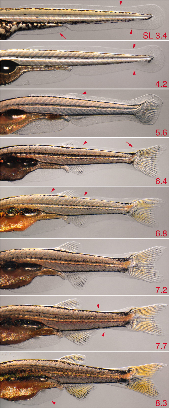
Fig. 23 Larval fin fold and fin fold resorption. Multiple individuals are shown (standard length [SL] at lower right). 3.4, Initial shapes of fin fold major lobe (arrowheads) and minor lobe (arrow); 4.2, a constriction is evident at the posterior tail (arrowhead); 5.6, a bulge is evident in the dorsal fin fold above the dorsal fin mesenchymal condensation (arrowhead); 6.4, a notch posterior to the dorsal fin indicates early fin fold resorption (arrowhead) and a bulge is evident over the developing supranotochordal fin rays (arrow); 6.8, resorption continues both anterior and posterior to the dorsal fin (arrowheads); 7.7, resorption occurs in an increasingly posterior zone along the tail (arrowheads); 8.3, early resorption of the minor lobe is revealed by flattening of its ventral posterior margin (arrowhead; also see Fig. 22).