
|
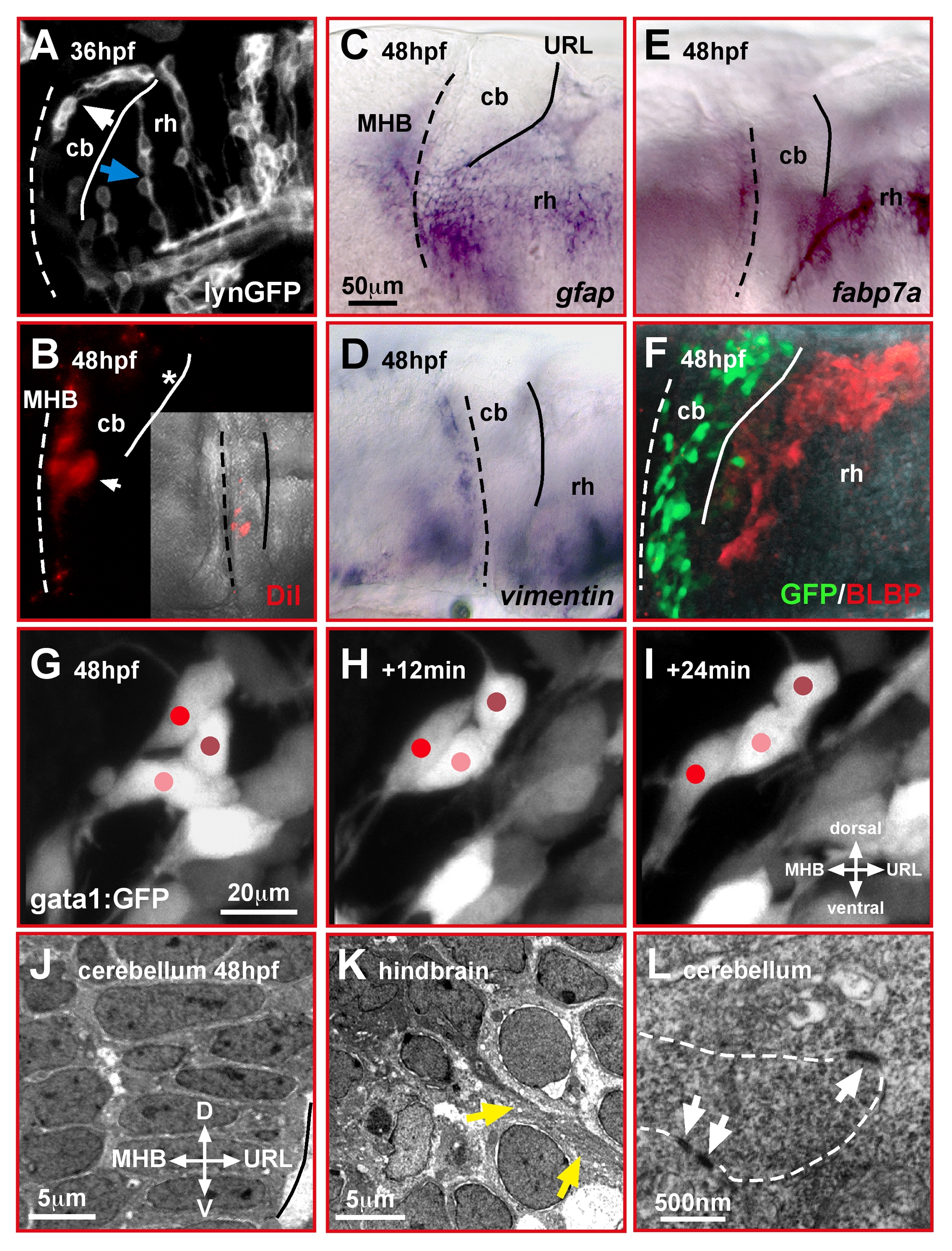
Fig. 1 Migrating cerebellar GCs interact in a homophilic manner.
(A–F) Lateral view of the zebrafish cerebellum and caudal hindbrain. Neither mosaic expression of membrane-targeted lynGFP (A) nor DiI-injection into the URL (B) reveals glia-like cell morphologies in the cerebellum, unlike that found in the hindbrain (blue arrow). In the cerebellum, GCs emigrating from the URL form chain-like structures (white arrow). Expression of the glia markers gfap (C), vimentin (D), fabp7a (E), and BLBP-immunohistochemistry in gata1:GFP embryos (F) supports that glia cells are absent in the cerebellum during GC migration. (G–I) In vivo time-lapse images (lateral view) of fluorescent GCs in gata1:GFP WT embryos reveals intense interactions among migrating GCs, which alternate in forward movements and resting behavior (see Video S1). (J–L) The differentiating cerebellum shows a highly ordered cellular arrangement when analyzed by TEM (J), with direct contacts of cerebellar cells via adherens junctions (L, white arrows). In the hindbrain (K), in contrast, fiber-like processes (yellow arrows) are present between individual cells (see also A, blue arrow). The MHB and URL are marked with a dashed and solid line, respectively. cb, cerebellum; MHB, midbrain-hindbrain boundary; rh, rhombencephalon.