
|
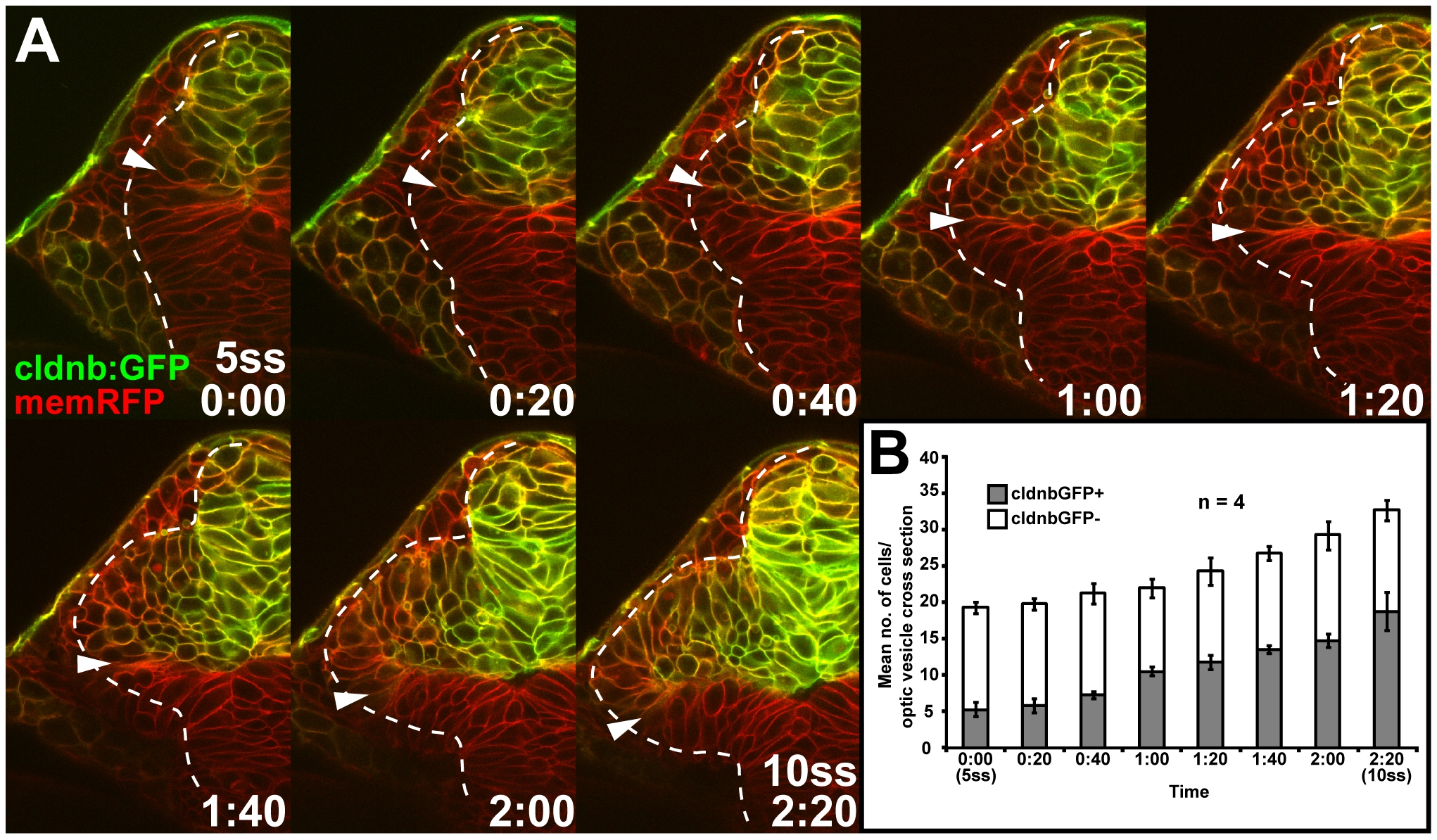
Fig. S3 In vivo imaging of clnb:GFP expression between the 5- and 10ss stages. (A) Single images from a confocal time-lapse series of cldnb:GFP expression (green) colabeled with membrane-targeted RFP (memRFP, red), captured at 20-min intervals (cross-section through one half of the forebrain, lateral to the left and dorsal to the top, bottom right: time in hours:minutes). (B) Mean number of cldnb:GFP-expressing (cldnbGFP+, grey) and nonexpressing (cldnbGFP-, white) cells in single cross-sections (captured at a 50–70-μm depth from anterior optic vesicle tip) through the optic vesicle between 5- and 10ss (error bar: standard deviation).