
|
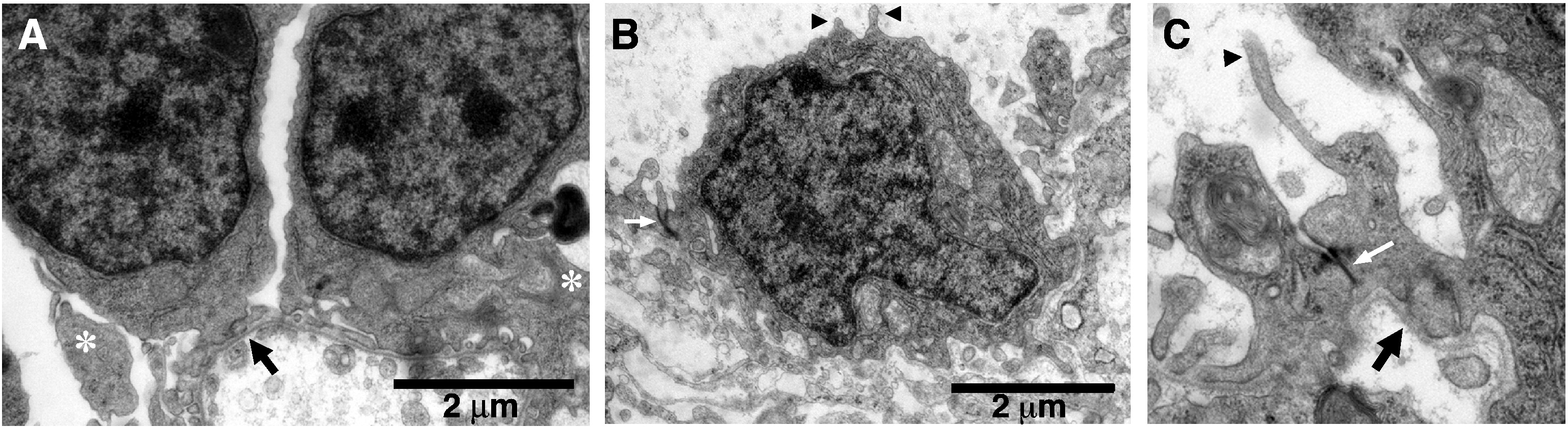
Fig. S2 Tight junctions persist in crb2b MO. (A) Electron micrographs of podocytes from control 4 dpf larvae shows the cell bodies, primary processes (asterisk), and the regular patterned array of foot processes along the GBM; GBM (black arrows). (B, C) In crb2b morphants, podocytes are attached to a GBM (black arrow in C), but the organization of regularly spaced foot processes is lost. Slit diaphragms are absent. Ectopic tight junctions are visible in morphant podocytes (white arrows in B and C). In addition, blunt ended apical membrane projections are found extending into the Bowman′s space (black arrowheads in B and C). A, B 20,000x magnification; C 40,000x magnification.
Reprinted from Developmental Biology, 334(1), Ebarasi, L., He, L., Hultenby, K., Takemoto, M., Betsholtz, C., Tryggvason, K., and Majumdar, A., A reverse genetic screen in the zebrafish identifies crb2b as a regulator of the glomerular filtration barrier, 1-9, Copyright (2009) with permission from Elsevier. Full text @ Dev. Biol.