
Image
|
Figure Caption
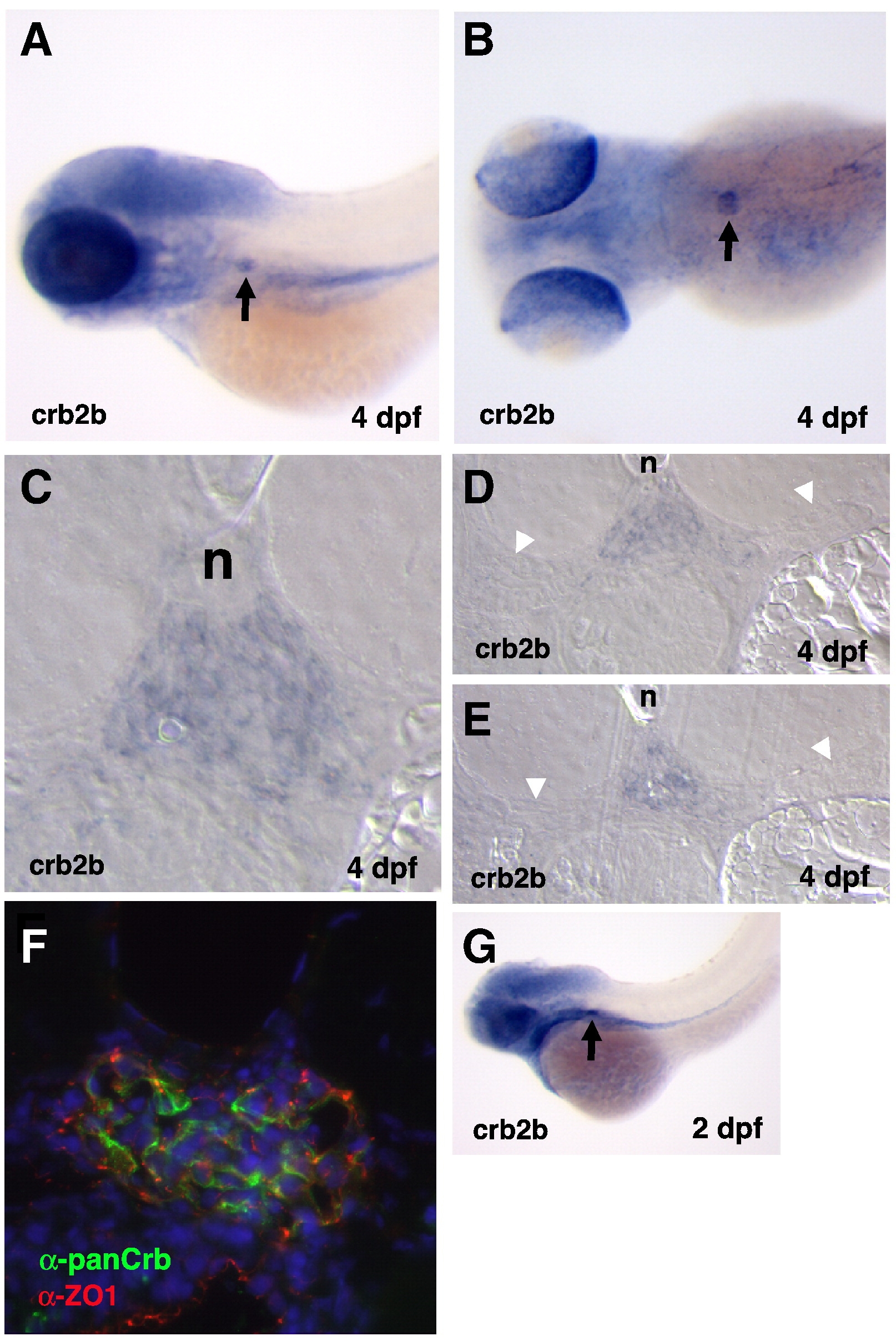
Fig. S1 crb2b expression in the pronephric glomerulus. (A, B) Wholemount in situ hybridization reveals crb2b expression in the glomerular podocytes and retina at 4 dpf and 2 dpf (G). Arrows point to the podocyte expression. (C, D, E) DIC images of transverse sections through a 4 dpf larval glomerulus stained with the crb2b antisense in situ probe. Notice that crb2b expression is not observed in the tubular epithelia (arrowheads; n, notochord). (F) Transverse section through a 4 dpf glomerulus double stained with α-panCrb (green) and α-ZO-1 (red) antibodies. Nuclei are stained with DAPI.
Acknowledgments
This image is the copyrighted work of the attributed author or publisher, and
ZFIN has permission only to display this image to its users.
Additional permissions should be obtained from the applicable author or publisher of the image.
Reprinted from Developmental Biology, 334(1), Ebarasi, L., He, L., Hultenby, K., Takemoto, M., Betsholtz, C., Tryggvason, K., and Majumdar, A., A reverse genetic screen in the zebrafish identifies crb2b as a regulator of the glomerular filtration barrier, 1-9, Copyright (2009) with permission from Elsevier. Full text @ Dev. Biol.