
|
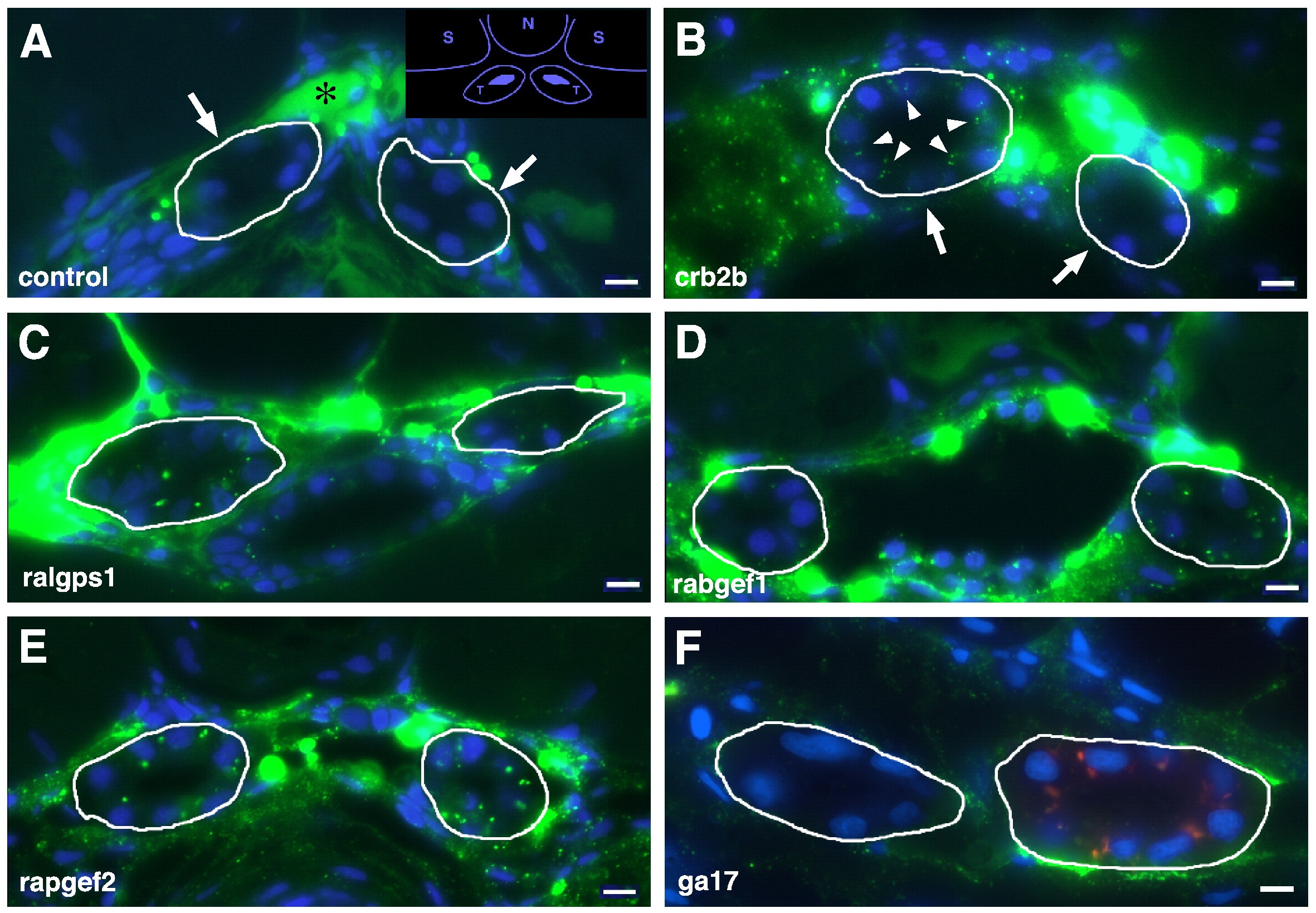
Fig. 1 Glomerular dye filtration assays on morphants. (A) Control morpholino injected 4 dpf larvae show no uptake of the 500 kDa FITC dextran dye into pronephric tubules (circled by white line). Dye is present within the dorsal aorta lumen (asterisk). In the inset, a schematic of a transverse section shows the relative positions of the somites (S), notochord (N), and pronephric tubules (T). (B) In crb2b morphants, the 500 kDa FITC dye is taken up into endosomes in the pronephric tubule epithelial cells (arrowheads). Similar uptake of the 500 kDa FITC dextran tracer is observed in ralgps1(C), rabgef1 (D), rapgef2 (E) morphants, but not in ga17 morphants (F), which tested negative in this assay. Nuclei are labeled with DAPI. Scale bar, 20 μm.
Reprinted from Developmental Biology, 334(1), Ebarasi, L., He, L., Hultenby, K., Takemoto, M., Betsholtz, C., Tryggvason, K., and Majumdar, A., A reverse genetic screen in the zebrafish identifies crb2b as a regulator of the glomerular filtration barrier, 1-9, Copyright (2009) with permission from Elsevier. Full text @ Dev. Biol.