
Image
|
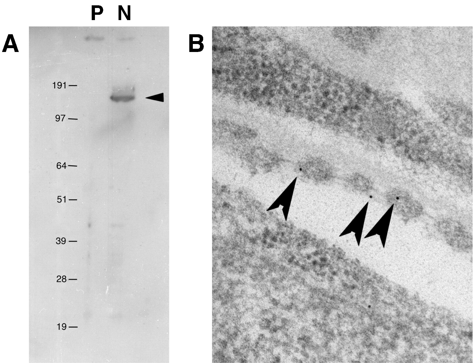
Figure Caption
Fig. S5 (A) Rabbit polyclonal antibodies directed against zebrafish Nephrin specifically recognize a single approximately protein in agreement with its predicted 140 ka size. Cell lysates from pcDNA3.1 (vector only, P) and zebrafish nephrin transfected HEK-293 cells, N. (B) Immuno-electron micrograph on wildtype podocytes with α-Nephrin antibody. Gold particles are found on the inner aspect of the slit diaphragms (arrowheads).
Acknowledgments
This image is the copyrighted work of the attributed author or publisher, and
ZFIN has permission only to display this image to its users.
Additional permissions should be obtained from the applicable author or publisher of the image.
Reprinted from Developmental Biology, 334(1), Ebarasi, L., He, L., Hultenby, K., Takemoto, M., Betsholtz, C., Tryggvason, K., and Majumdar, A., A reverse genetic screen in the zebrafish identifies crb2b as a regulator of the glomerular filtration barrier, 1-9, Copyright (2009) with permission from Elsevier. Full text @ Dev. Biol.