
|
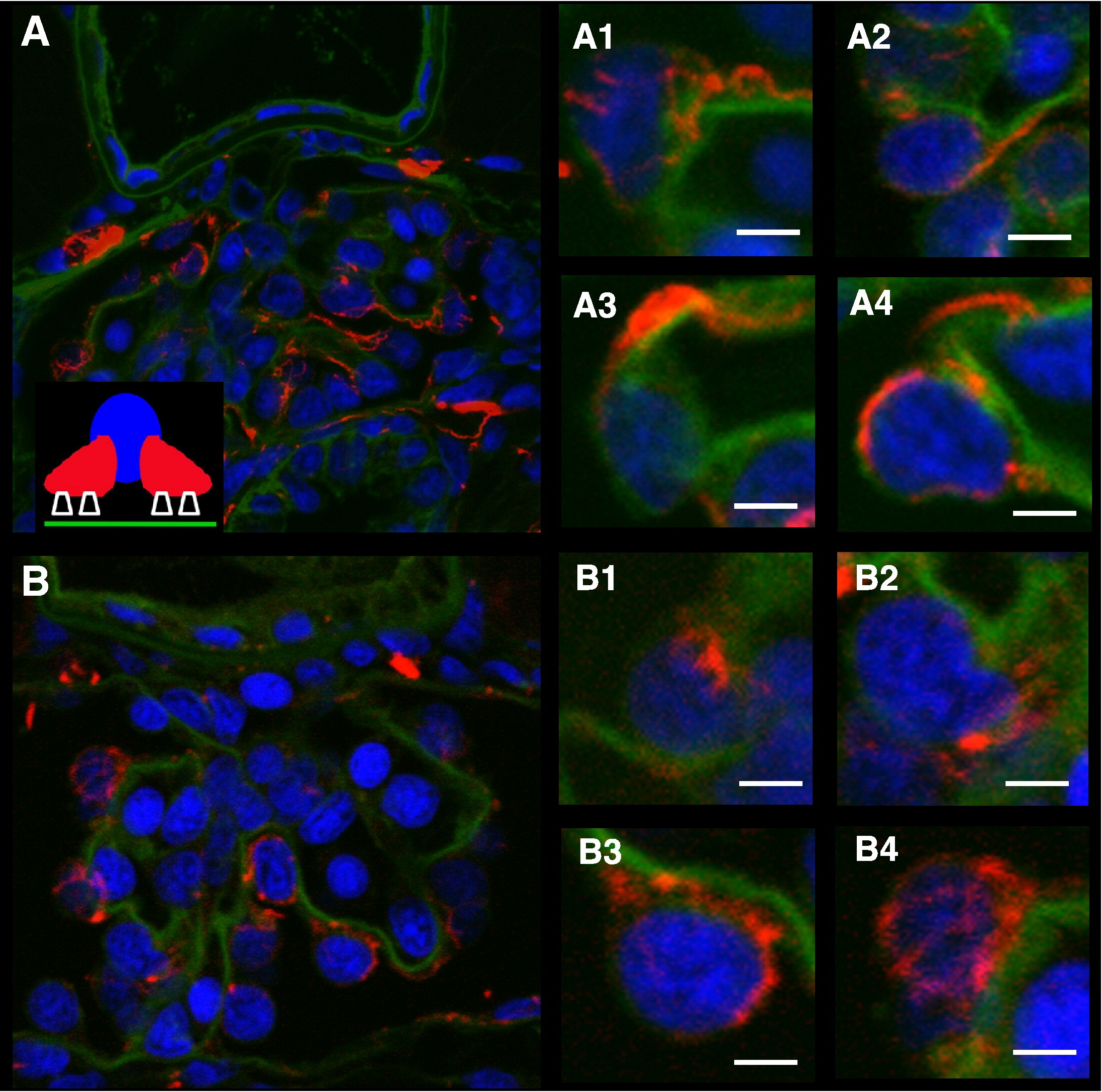
Fig. S4 Overall polarity is maintained in crb2b MO podocytes. Immunofluorescence staining of 4 dpf control (A, transverse glomerular section and A1–A4, individual podocytes) and crb2b MO (B, transverse glomerular section and B1–B4, individual podocytes) glomeruli with α-acetylated Tubulin, which labels the microtubule rich podocyte primary processes. Individual podocytes are chosen as examples. Primary processes are found lateral and basal to the podocyte nuclei, but apical to the GBM. Podocytes are identified by virtue of their unique position on the outer aspect of the capillary loops with their cell bodies in the Bowman′s space. The inset figure in A schematizes the relative positions of nucleus (blue), primary processes (red), foot processes (not stained, white), and GBM (green). Nuclei stained with DAPI. GBM stained with WGA FITC. Scale bars, 2 μM.
Reprinted from Developmental Biology, 334(1), Ebarasi, L., He, L., Hultenby, K., Takemoto, M., Betsholtz, C., Tryggvason, K., and Majumdar, A., A reverse genetic screen in the zebrafish identifies crb2b as a regulator of the glomerular filtration barrier, 1-9, Copyright (2009) with permission from Elsevier. Full text @ Dev. Biol.