
|
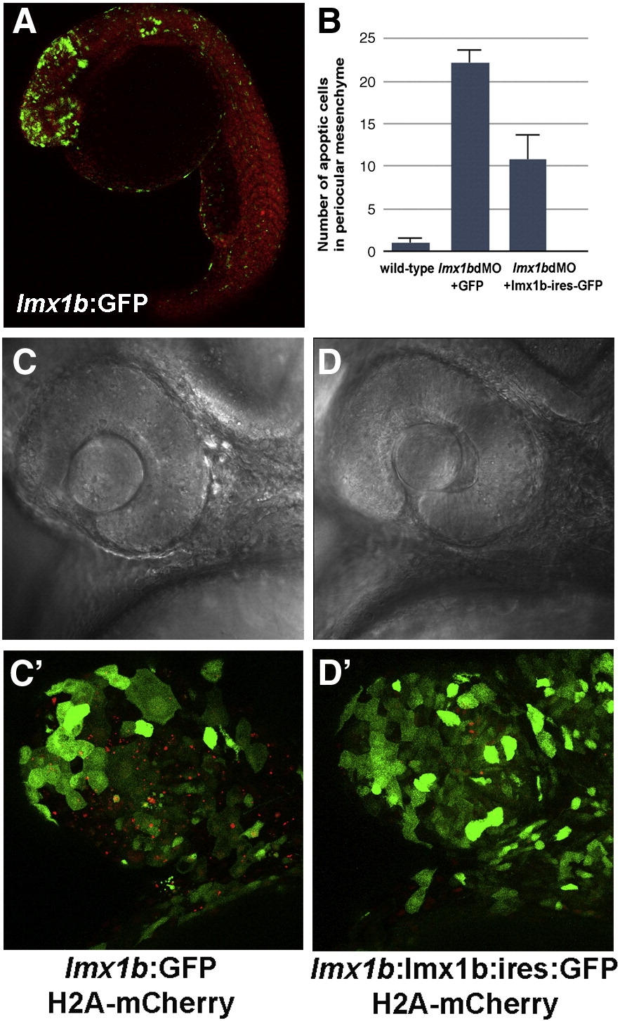
Fig. 4 Apoptosis in lmx1bdMO morphants is rescued by expression of lmx1b.1 cDNA. (A) Expression of GFP driven by the - 5 kb lmx1b.1 promoter. (B) Quantitation of the number of pyknotic cells present in ocular regions from wild-type, lmx1bdMO morphants expressing GFP, and lmx1bdMO embryos expressing lmx1b.1:ires:GFP. For each condition, n = 10 independent eyes were scored. Error bars represent Standard Error of the Mean. (C) Bright field image of lmx1bdMO morphants expressing GFP or (D) lmx1bdMO morphants expressing lmx1b.1:ires:GFP. (C′) Fluorescent image of panel C showing lmx1b:GFP-positive periocular cells (green) and pyknotic cells red). (D′) Fluorescent image of panel D showing lmx1b:lmx1b.1:ires:GFP-positive periocular cells (green) and pyknotic periocular cells (red).
Reprinted from Developmental Biology, 332(2), McMahon, C., Gestri, G., Wilson, S.W., and Link, B.A., Lmx1b is essential for survival of periocular mesenchymal cells and influences Fgf-mediated retinal patterning in zebrafish, 287-298, Copyright (2009) with permission from Elsevier. Full text @ Dev. Biol.