
|
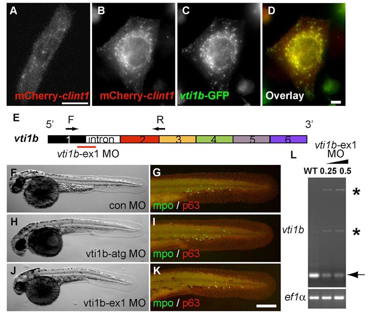
Fig. S4 The zebrafish proteins Clint1 and Vti1b co-localize but have non-overlapping roles in epidermal development. (A-D) Expression of mCherry-clint1 in zebrafish epidermal cell (A) or mCherry-clint1 (B,D) and vti1b-GFP (C,D) in HEK293 cells. (E) Schematic of vti1b exon structure and primers used to investigate effects of vti1b-ex1 MO on splicing. (F-K) Control MO (F,G), vti1b-atg MO (H,I) or vti1b-ex1 MO (J,K) injected embryos immunolabeled for MPO (green) and p63 (red). (L) RT-PCR amplification of vti1b and ef1a from control and clint1-ex1 MO-injected wild-type embryos. Arrow and asterisks indicate full-length and alternatively spliced vti1b transcripts, respectively. Scale bars: 10 μm in A,D; 200 μm in K.