
|
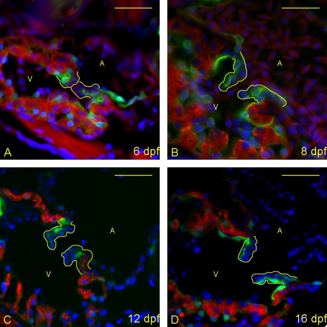
Fig. 1 Zebrafish heart valves elongate but remain highly cellular between 6 and 16 days postfertilization (dpf). A-D: Five micrometer paraffin sections of flk1::GFP (GFL, green fluorescent protein) transgene-carrying zebrafish embryos/larvae were imaged to identify the morphology of the atrioventricular (AV) boundary and intraluminal structure. Atria (A) and ventricles (V) are labeled (yellow letters), and yellow lines outline the extent of the AV structure (cushion and/or valve). Staining identifies cell nuclei (DAPI, 4′,6-diamidine-2-phenylidole-dihydrochloride; blue), myocardium (anti-MF-20: red), and endothelial cells (anti-GFP; green). Through this time period, the valves progressively lengthen and thin, and little to no extracellular matrix is seen within the valve leaflets. The most significant change is in cellular organization from 12 (C) to 16 (D) dpf, with the leaflets developing a linear, two-cell-layer thick form. Scale bar = 20 μm.