
|
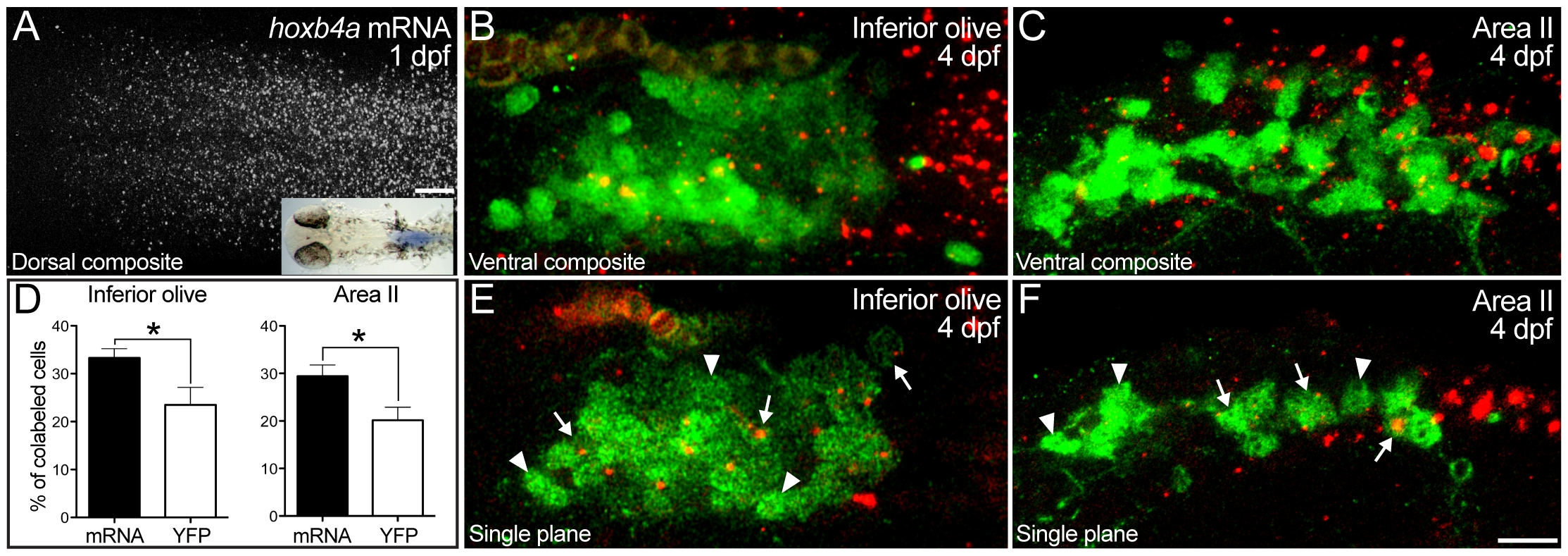
Fig. 2 Mosaic hoxb4a expression within hindbrain nuclei.
(A) Hoxb4a expression in hindbrain r7–8 of a 1 dpf larva detected by fluorescent and colormetric procedures (inset). (B–C) Ventral composites of the precerebellar inferior olive (B) and Area II (C) nuclei (20 μm confocal stacks) retrogradely labeled from the cerebellum at 4 dpf (green), with hoxb4a mRNA (red) detected by fluorescent in situ hybridization. (D) Graphs showing only a subset of inferior olive and Area II neurons expressed hoxb4a mRNA (solid bar) and hoxb4a-YFP (open bar). P-values from Student t-test were 0.04 for both the inferior olive and Area II. (E–F) Single plane images showing mosaic hoxb4a expression in the olive (E) and Area II (F). Arrows and arrowheads point to cells that did and did not expression hoxb4a. Scale bar = 20 μm (A) and 10 μm (B–C and E–F).