
|
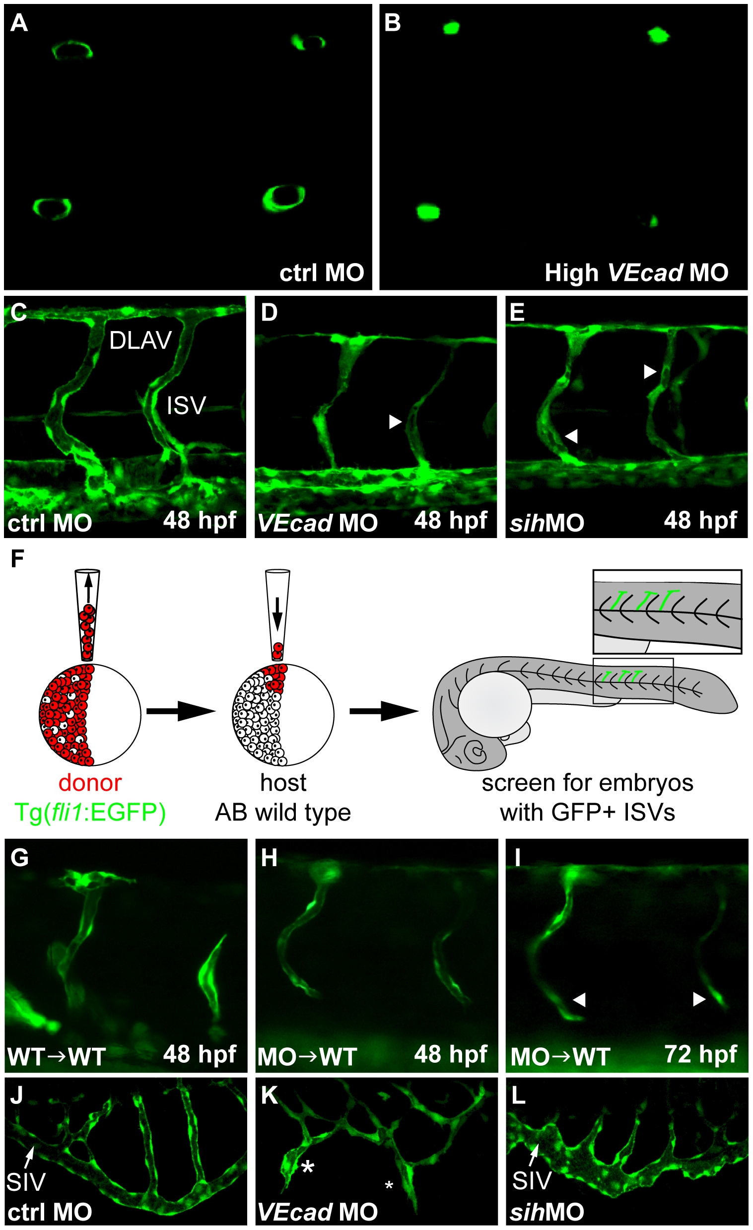
Fig. 6 VE-cadherin expression influences lumen formation.
(A–B) Optical cross sections of intersegmental vessels (ISVs) at 48 hpf in Tg(fli1:EGFP) background acquired at the confocal microscope by positioning the embryo on a dorsal orientation. CtrlMO injected embryos have fully lumenized ISVs (A) while VE-cadherin severe morphants (High VEcadMO) do not (B). (C–E) Lumenization of ISVs is influenced by blood circulation. Lateral views of trunk vessels of ctrlMO (C), high VEcadMO (D) and sihMO (E) injected embryos at 48 hpf. At this stage, ISVs and the dorsal longitudinal anastomotic vessels (DLAV) are fully lumenized in control embryos. In contrast, VE-cadherin and sih morphants present only a very small lumen in some portions of the ISVs (arrowheads in D and E) and essentially no lumen in DLAV. (F–I) Transplantation experiments. (F) Scheme of the experimental procedure. Donor cells from fli1:EGFP embryos were transplanted into AB wild type embryos at 4 hpf and, after development, embryos with EGFP positive ISVs were selected for analysis. When wild type embryos were used as donor (G), ISVs did form lumen and were integrated into the host vasculature at 48 hpf. Morphant donor cells transplanted into wild type embryos form thin lumens at 48 hpf (H) that fail to correctly integrate in the host vasculature and possibly collapse at 72 hpf (arrowheads in panel I). (J–L) Lumenization of subintestinal vein (SIV) is independent of blood flow as it occurs in the absence of heart beat in sih morphants at 72 hpf (L) but is dramatically affected by the absence of VE-cadherin. High VEcadMO SIVs display only partially lumenized regions which are not fully interconnected (asterisks in panel K).