
|
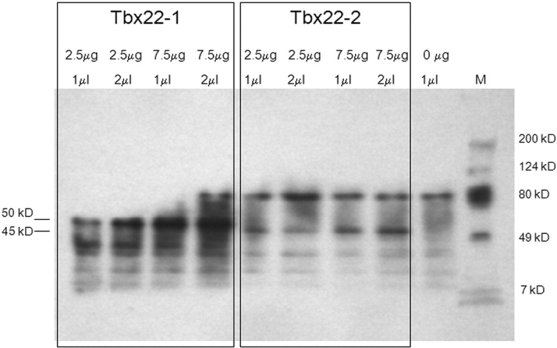
Fig. 3 In vitro transcription and translation of tbx22 splice variants. Western blot analysis revealed distinct biotinylated Tbx22-1 and Tbx22-2 in vitro translation products corresponding to the predicted 50.3- and 45.2-kD Tbx22 isoforms, respectively. No putative 48.3-kD Tbx22-1 translation product was observed, consistent with the location of the Tbx22-1 start codon at bp 147. Plasmid concentrations used in the in vitro transcription reactions, and the amount of translation product loaded in each well, are as indicated. A no-template negative control is also shown. M, Molecular weight protein standard markers.