
|
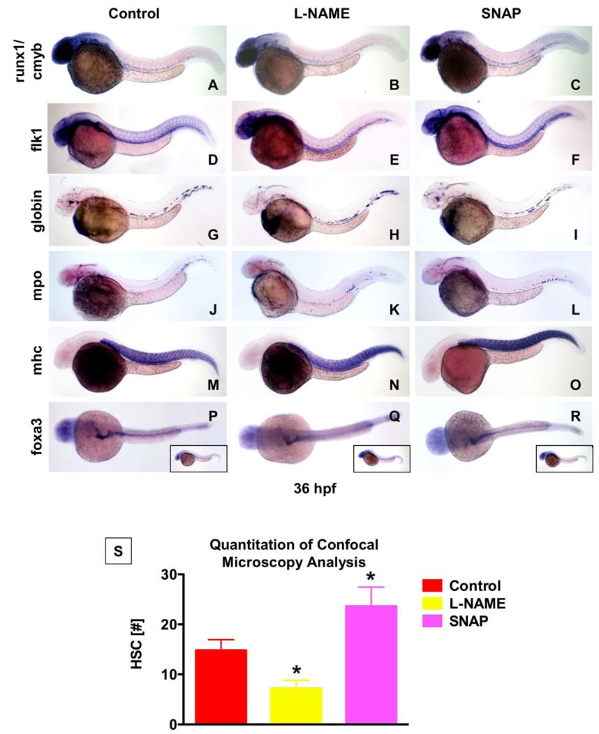
Fig. S5
NO modulation does not affect primitive hematopoiesis or development of mesodermal and endodermal structures.
(A-R) Embryos (n>25 per treatment) were exposed to control, L-NAME or SNAP from 10 somites to 36 hpf.
(A-F) Expression of the vascular marker flk1 is minimally altered by NO modulation.
(G-R) Primitive erythro- (globin) and myelopoiesis (mpo) as well as early muscle (mhc) and endoderm (foxa3) development are not affected by changes in NO signaling.
(S) Quantitation of HSC number in confocal microscopy analysis of cmyb:GFP; lmo2:dsRed embryos (Fig.3Q-S) reveals significant changes in response to NO modulation (* sig vs. control, ANOVA, p<0.001, n=5).
Reprinted from Cell, 137(4), North, T.E., Goessling, W., Peeters, M., Li, P., Ceol, C., Lord, A.M., Weber, G.J., Harris, J., Cutting, C.C., Huang, P., Dzierzak, E., and Zon, L.I., Hematopoietic stem cell development is dependent on blood flow, 736-748, Copyright (2009) with permission from Elsevier. Full text @ Cell