
|
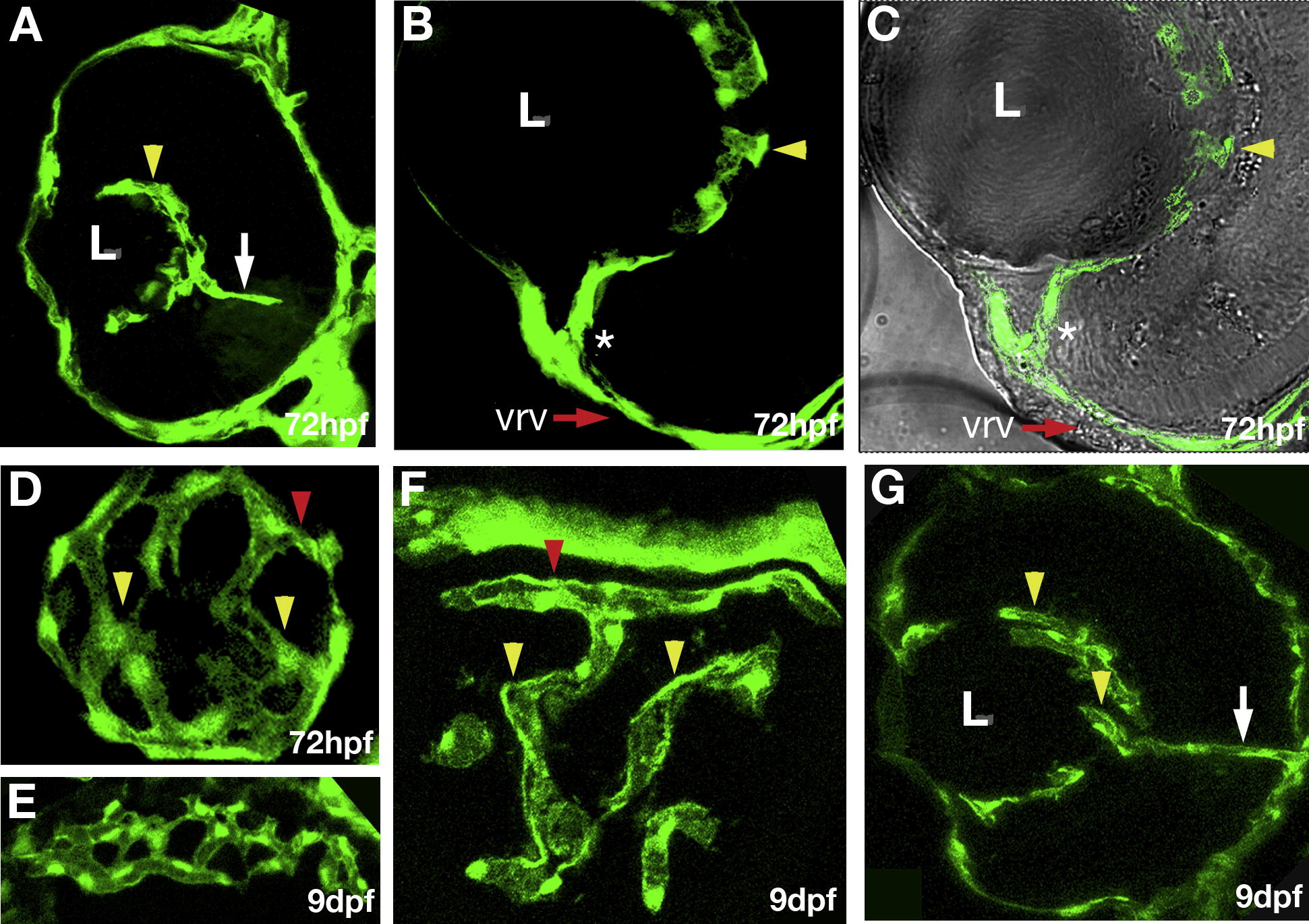
Fig. 2 Characterization of early retinal vasculature. Confocal images of cryosections through eyes of fli1:EGFP transgenic animals. (A–C) Transverse sections through the retina. Intraocular vessels (yellow arrowheads) wrap around the lens, and are connected to the surface vessel (asterisk) at 72 hpf. Dorsal is up and lateral to the left. (D) A somewhat oblique longitudinal section through the region behind the lens reveals a vessel network on the surface of the lens. Posterior is to the right, lateral is down. (E) A transverse tangential section through the rostral eye showing the choriodal vessel network at 9 dpf. (F) A transverse section tangential to the lens showing the intraocular ring vessel (red arrowhead) and the intraocular vessel network (yellow arrowheads) at 9 dpf. (G) A transverse section showing intraocular vessels at 9 dpf, dorsal is up, and lateral to the left. The retinal artery is indicated by white arrow.
Reprinted from Mechanisms of Development, 126(5-6), Kitambi, S.S., McCulloch, K.J., Peterson, R.T., and Malicki, J.J., Small molecule screen for compounds that affect vascular development in the zebrafish retina, 464-477, Copyright (2009) with permission from Elsevier. Full text @ Mech. Dev.