
Image
|
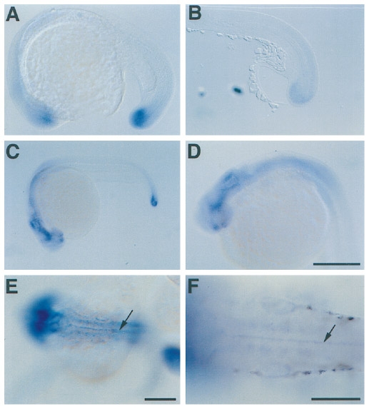
Figure Caption
Fig. 6 Whole-mount in situ hybridization of Cx43.4 mRNA during late segmentation and pharyngula periods. (A) 20-somite stage embryo; side view with anterior to the left. (B) Sagital section through the tail at the 17-somite stage. (C and D) End of segmentation period; side view with anterior to the left. (E and F) Pharyngula period (30 hr); dorsal view with anterior to the left. Arrows point to the floor plate of the central nervous system. Scale bars: D, 250 μm, and the magnification is the same for A and B; E, 250 μm, and the same for C; F, 100 μm.
Acknowledgments
This image is the copyrighted work of the attributed author or publisher, and
ZFIN has permission only to display this image to its users.
Additional permissions should be obtained from the applicable author or publisher of the image.
Reprinted from Developmental Biology, 177(2), Essner, J.J., Laing, J.G., Beyer, E.C., Johnson, R.G., and Hackett, P.B. Jr., Expression of zebrafish connexin43.4 in the notochord and tail bud of wild-type and mutant no tail embryos, 449-462, Copyright (1996) with permission from Elsevier. Full text @ Dev. Biol.