
|
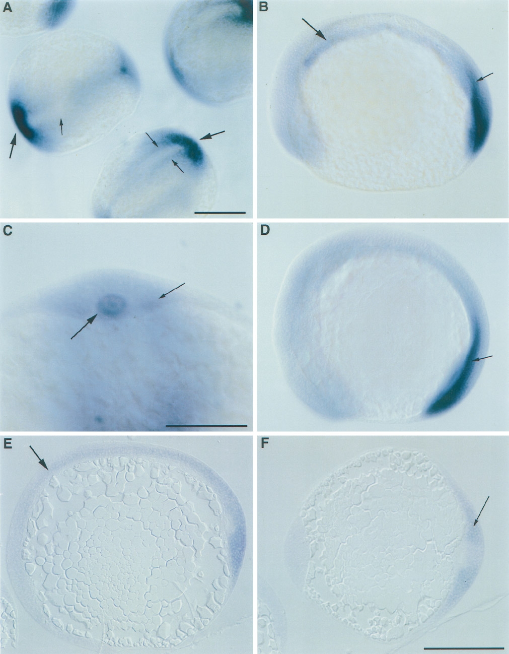
Fig. 5 Whole-mount in situ hybridization of Cx43.4 mRNA during segmentation. (A) 2- to 5-somite stage; dorsal views. Large arrow, tail bud; small arrows, paraxial mesoderm. Scale bar, 250 μm. (B) 2-somite stage; side view. Large arrow, notochord; small arrow, paraxial mesoderm. (C) 5-somite stage; posterior, transverse view at higher magnification. Large arrow, notochord; small arrow, paraxial mesoderm. Scale bar, 100 μm. (D) 10-somite stage, side view, anterior to the left; arrow points to paraxial tissue. (E) Sagital section of a 1-somite embryo; arrow marks notochord. (F) Transverse section of a 2-somite stage embryo, arrow marks expression in paraxial tissue in the tail. Scale bar in F represents 250 μm and is the same for B, D, and E.
Reprinted from Developmental Biology, 177(2), Essner, J.J., Laing, J.G., Beyer, E.C., Johnson, R.G., and Hackett, P.B. Jr., Expression of zebrafish connexin43.4 in the notochord and tail bud of wild-type and mutant no tail embryos, 449-462, Copyright (1996) with permission from Elsevier. Full text @ Dev. Biol.