
|
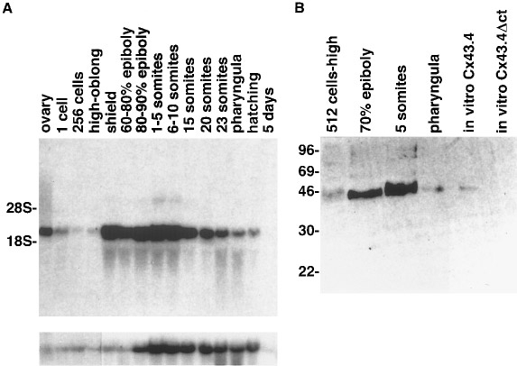
Fig. 3 Temporal expression of Cx43.4. (A) Upper panel: Northern blot hybridized with an antisense Cx43.4 RNA probe. 10 μg of total RNA was loaded in each lane for each developmental stage indicated. The mobilities of the 18S (2.1 kb) and the 28S (5.2 kb) rRNAs are indicated at the side. Lower panel: The same membrane was rehybridized with an antisense eF-1α RNA probe as a control for integrity of the mRNA. (B) Western blot of immunoprecipitated Cx43.4 protein from embryos. The first four lanes represent 10 embryos from each stage. The lanes labeled in vitro Cx43.4 and Cx43.4Δct involve immunoprecipitations of proteins translated in vitro from Cx43.4 mRNA and a mutant Cx43.4 mRNA lacking most of the anti-Cx45 epitope region, respectively.
Reprinted from Developmental Biology, 177(2), Essner, J.J., Laing, J.G., Beyer, E.C., Johnson, R.G., and Hackett, P.B. Jr., Expression of zebrafish connexin43.4 in the notochord and tail bud of wild-type and mutant no tail embryos, 449-462, Copyright (1996) with permission from Elsevier. Full text @ Dev. Biol.