
|
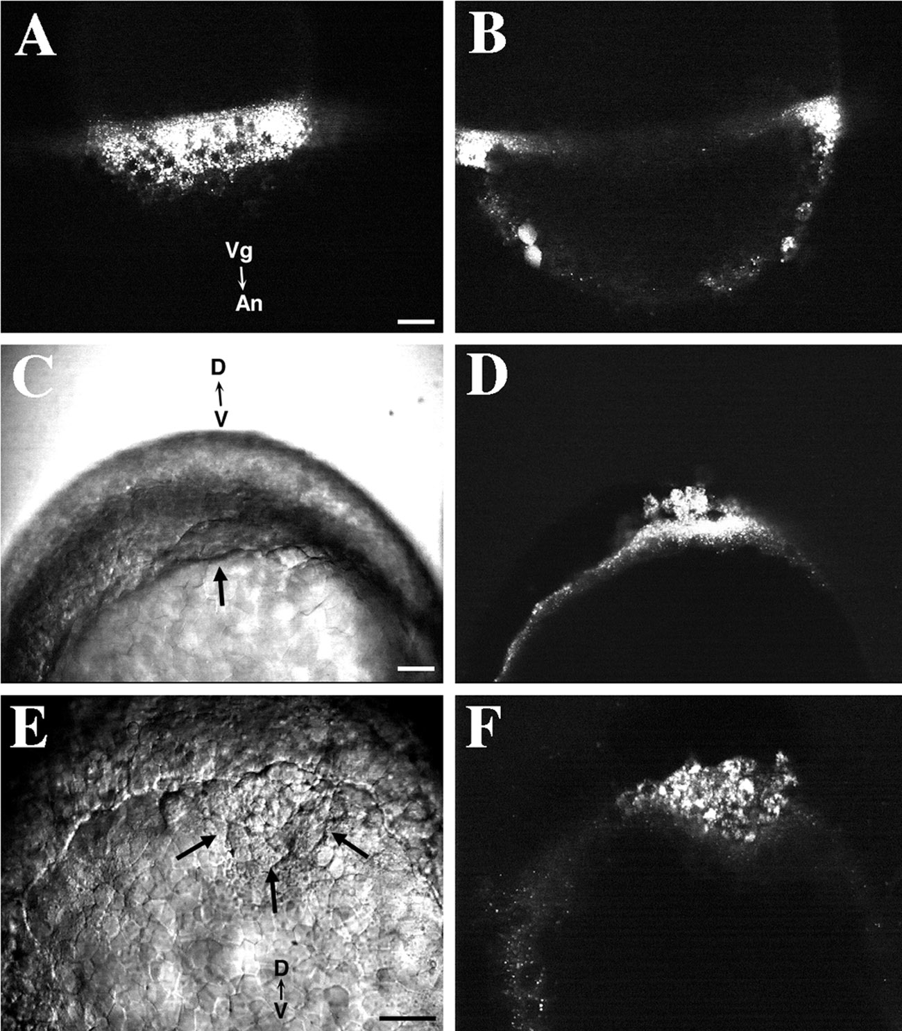
Fig. 6 Membrane impermeant fluorescent dyes are endocytosed into the YSL and cytoplasm of NEM cells. Scale bars, 50 μm. (A) FITC-labeled dextran (10K MW) is internalized by fluid-phase endocytosis into the YSL. Numerous endosomes are distributed through the YSL. Nuclei in the YSL are unlabeled. A confocal view of the EVL–YSL margin. The embryo was labeled between oblong and sphere stage and observed at 30%-epiboly. (B) A confocal section deeper into the same embryo as shown in A. No labeling of deep cells is seen. Two EVL cells are diffusely labeled with dextran (bottom left). (C) A Nomarski view of a zebrafish embryo at 70%-epiboly. The embryo was labeled with dextran at 50%-epiboly. The characteristic thickening at the dorsal margin of the blastoderm is visible (arrow). View of the embryo from the vegetal pole. (D) Fluorescence confocal image of the same embryo as in C. Numerous fluorescently labeled endosomes are visible in the cytoplasm of the YSL and closely associated forerunner cells. (E) A Nomarski image of the blastoderm edge of an embryo, viewed from the vegetal pole, at 80%-epiboly. The forerunner cell cluster (arrow) is visible as a wedge of cells located distal to the advancing dorsal margin of the blastoderm. The embryo was labeled with fluorescent dextran at 50%-epiboly. (F) Fluorescence confocal image of the same area as shown in E. Forerunner cells possess numerous endosomes containing FITC–dextran. A sparser distribution of fluorescently labeled endosomes is visible within the YSL in the left portion of the image.
Reprinted from Developmental Biology, 180(1), Cooper, M.S. and D'Amico, L.A., A cluster of noninvoluting endocytic cells at the margin of the zebrafish blastoderm marks the site of embryonic shield formation, 184-198, Copyright (1996) with permission from Elsevier. Full text @ Dev. Biol.