
|
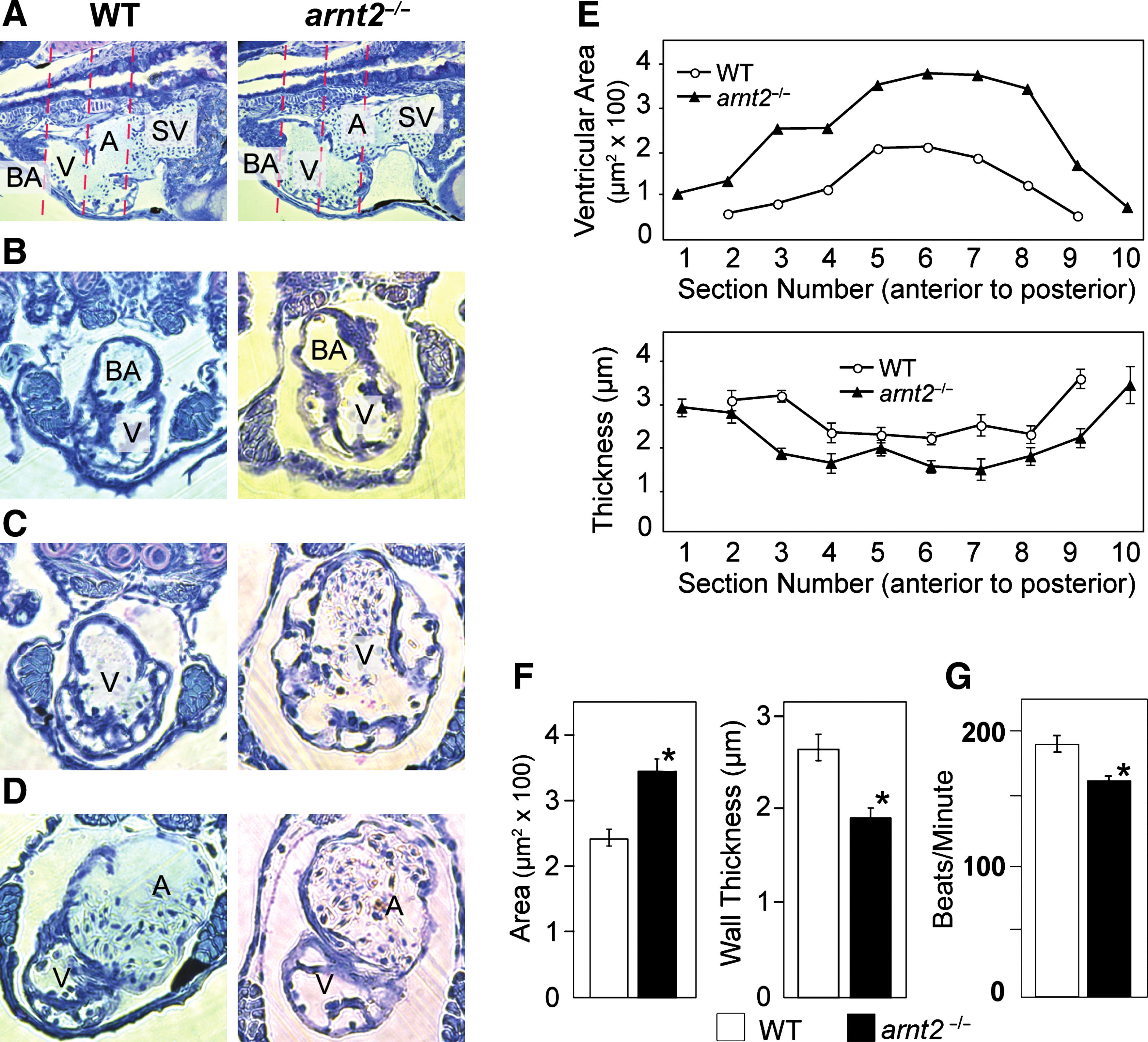
Fig. 8 Heart ventricle enlargement, bradycardia, and cardiac arrhythmia in arnt2-/- larvae at 120 and 144–148 hpf. In a representative longitudinal section of the heart (A) of a WT larva (left) and an arnt2-/- larva (right) at 120 hpf, the ventricle of the arnt2-/- mutant appears slightly larger than WT. Transverse sections of the ventricle at 120 hpf were taken along its anterior–posterior axis: anterior (B), middle (C), and posterior (D) for WT (left) and arnt2-/- mutant (right), respectively. A, Atrium; BA, bulbous arteriosus; SV, sinus venosus; V, ventricle. Line graphs (E) show ventricle cross-sectional area as a function of section number (top line graph) and ventricle wall thickness as a function of section number (bottom line graph). WT (line, open circle) and arnt2-/- (line, solid triangle) results shown in the line graphs are for n=7 larvae=genotype at 120 hpf. Mean ventricle area and ventricle wall thickness based on all cross section measurements of the ventricle also are shown for WT and arnt2-/- mutant’s heart (F, bar graphs, right and middle). Heart rate assessed in WT and arnt2-/- larvae at 144 hpf (G) revealed the presence of bradycardia in arnt2-/- larvae (WT, n=4; arnt2-/-, n=11). Asterisk indicates a significant difference (p<0.05).