
|
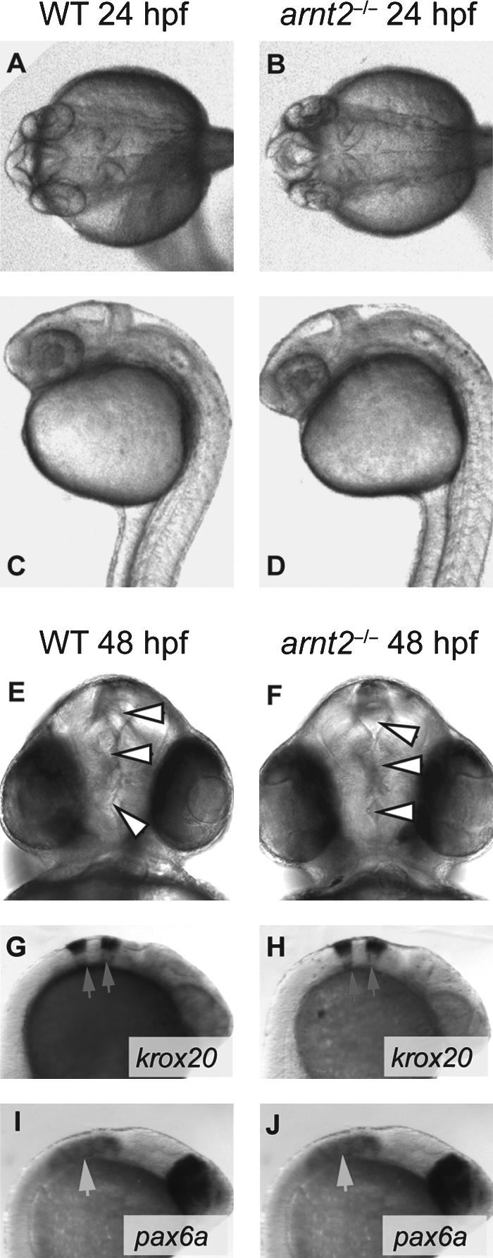
Fig. 3 Early brain development appears normal in arnt2-/- embryos at 24–48 hpf. Brain development in the arnt2-/- mutant embryo was assessed by gross examination of the brain at the light microscopic level at 24 hpf. A dorsal view of the developing brain of a WT embryo and an arnt2-/- embryo (A, B) and a lateral view of the brain of a WT embryo and an arnt2-/- embryo (C, D) are shown. At 48 hpf a dorsal view of the brain of a representative WT embryo and arnt2-/- embryo (E, F) is shown. White arrowheads point to ventricles in the brain of the WT and arnt2-/- mutant, demonstrating that brain ventricles are similar in size and shape in the two genotypes (E, F). Overall, gross brain morphology of arnt2-/- larvae at 24–48 hpf was indistinguishable from WT. Patterns of staining at 24 hpf are shown for whole-mount ISH of krox20 in a representative WT embryo and arnt2-/- embryo at 24 hpf (G, H, arrowheads) and of pax6a (I, J, arrowheads).