
|
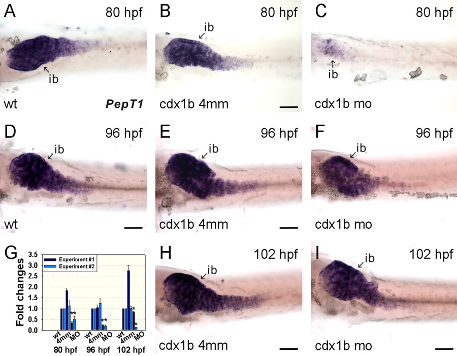
Fig. 5 Knockdown of cdx1b function affects PepT1 expression in enterocytes. A-C: Wild type (A), cdx1b-4mm MO-injected (B), and cdx1b MO-injected (C) 80-hr post-fertilization (hpf) embryos hybridized with PepT1 antisense RNA probes. D-F: Wild type (D), cdx1b-4mm MO-injected (E), and cdx1b MO-injected (F) 96-hpf embryos hybridized with PepT1 antisense RNA probes. G: Q-PCR indicated decreased PepT1 expression in cdx1b morphants compared to wild type and cdx1b-4mm MO-injected embryos from 80 to 102 hr of development. Error bars indicate standard errors. Results from duplicated experiments are shown. Significant differences between ΔΔCt values based on Student's t-test were used to indicate different gene expressions expressed as fold changes. Student's t-test was conducted to compare cdx1b MO-injected embryos with either wild type or cdx1b-4mm MO-injected embryos.*P < 0.05 in both comparisons. H, I: cdx1b-4mm MO-injected (H) and cdx1b MO-injected (I) 102-hpf embryos hybridized with PepT1 antisense RNA probes. Ib, intestinal bulb. Scale bars = 100 μm.