
|
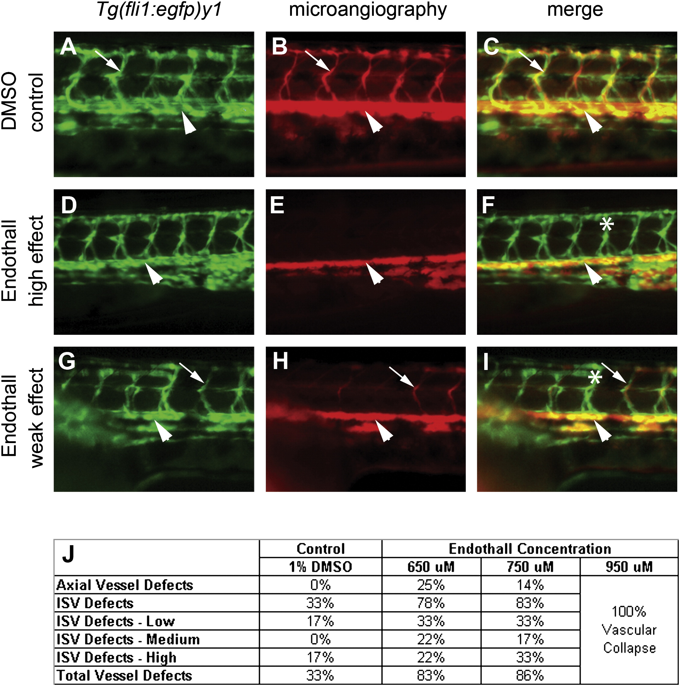
Fig. 5 Treatment of Embryos with Endothall Results in Defects in Tubulogenesis
(A, D, and G) Endothelial cells in the trunk were analyzed for defects in migration in the Tg(fli1:egfp)y1 line (green fluorescence) at 48–50 hpf. (B, E, and H) Analysis of circulation defects by using microangiography (rhodamine-dextran imaging, red) at 48–50 hpf. (A–I) are lateral views of the trunk. (A–C) Treatment with either carrier (DMSO control) or (D–I) Endothall did not alter endothelial migration (see Tg(fli1:egfp)y1 in [A], [D], and [G]). (B, E, and H) Microangiography revealed defects is circulation consistent with defects in tubulogenesis. Two classes of affected embryos are shown. In weakly affected embryos (low effect), some of the ISVs would fail to circulate rhodamine-dextran. In more severely affected embryos (high effect), most of the ISVs failed to transfer dye. (C, F, and I) Merged images of the previous two panels. Dorsal aortas are marked with arrowheads, examples of perfused ISVs are marked with arrows, and examples of non-perfused ISVs are labeled with asterisks. (J) Dose response to bathing embryos in Endothall. Embryos were scored based on circulation in the axial vessels and the ISVs as in Table 2. Vascular collapse was observed with the highest concentration of Endothall.