
|
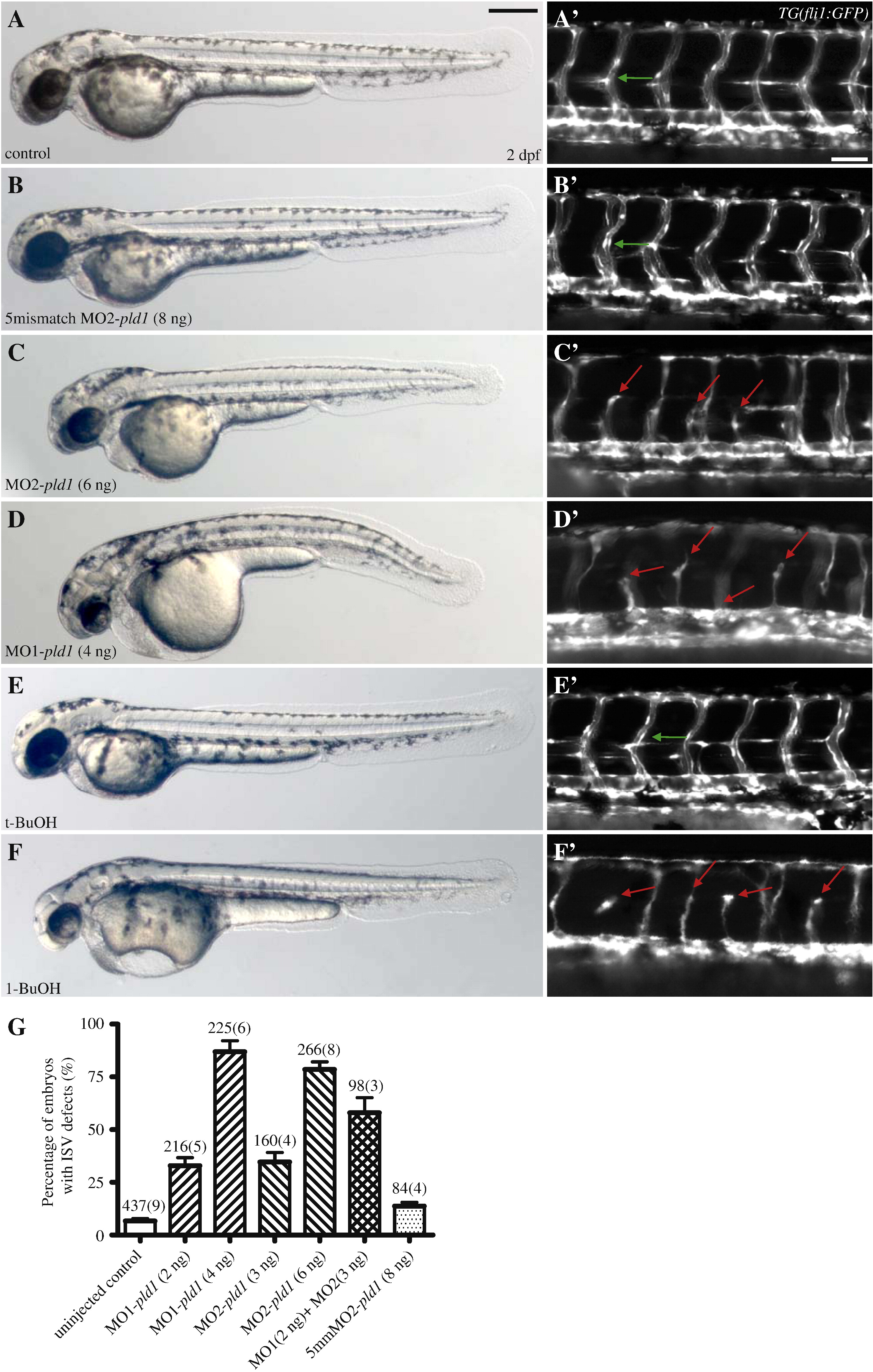
Fig. 4 Disruption of Pld1 signaling impairs development of intersegmental vessels. (A,B,C,D,E,F) Nomarski and (A′,B′,C′,D′,E′,F′) fluorescent images of trunk region of TG(fli1:GFP) embryos at 2 dpf. (A,A′) Uninjected control embryos, (B,B′) embryos injected with 8 ng 5mismatch MO2-pld1 and (C,C′) embryos injected with 6 ng MO2-pld1, or (D,D′) 4 ng MO1-pld1, (E,E′) 0.3% t-butanol treated embryo and (F,F′) to 0.3% 1-butanol treated embryo. (A′,B′,E′) Green arrows point to ISVs with normal morphology, whereas (C′,D′,F′) red arrows point to ISV defects, where ISVs stop in the middle along the ventral–dorsal axis or show completely blocked sprouting from the ventral blood vessels (D′,F′). Scale bar represents 20 μm (A′) and 250 μm (A). (G) A bar graph shows the percentage of ISV defects in embryos that were injected with different morpholinos. The total number of embryos is indicated above the bar as well as the number of experimental repeats (in parentheses).
Reprinted from Developmental Biology, 328(2), Zeng, X.X., Zheng, X., Xiang, Y., Cho, H.P., Jessen, J.R., Zhong, T.P., Solnica-Krezel, L., and Brown, H.A., Phospholipase D1 is required for angiogenesis of intersegmental blood vessels in zebrafish, 363-376, Copyright (2009) with permission from Elsevier. Full text @ Dev. Biol.