
|
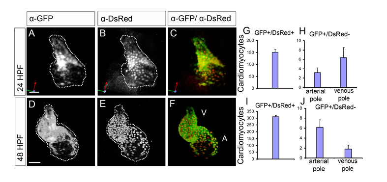
Fig. S3 eGFP and DsRed protein expression overlaps at 24 hpf and 48 hpf. (A-F) Reconstruction of confocal z-stacks of Tg(cmlc2:eGFP)/Tg(cmlc2:DsRed) embryos at 24 hpf and 48 hpf. Hearts stained immunohistochemically with α-eGFP (A,D) and α-DsRed (B,E) antibodies. C and F represent the overlay. At 24 hpf and at 48 hpf, both eGFP and DsRed protein expression overlaps throughout the heart, with the exception of a few cells. (G-J) The number of cells in Tg(cmlc2:eGFP)/Tg(cmlc2:DsRed) hearts that are eGFPposDsRedpos (G,I) or eGFPposDsRedneg (H,J; mean±s.e.m., n_5). The eGFPposDsRedneg cardiomyocytes are subdivided into those present at the arterial pole and those present at the venous pole (24 hpf, 3±1 and 6±2; 48 hpf, 6±1 and 2±1, respectively). Note that almost all cardiomyocytes are eGFPposDsRedpos (24 hpf, 151±12; 48 hpf, 311±10).