
|
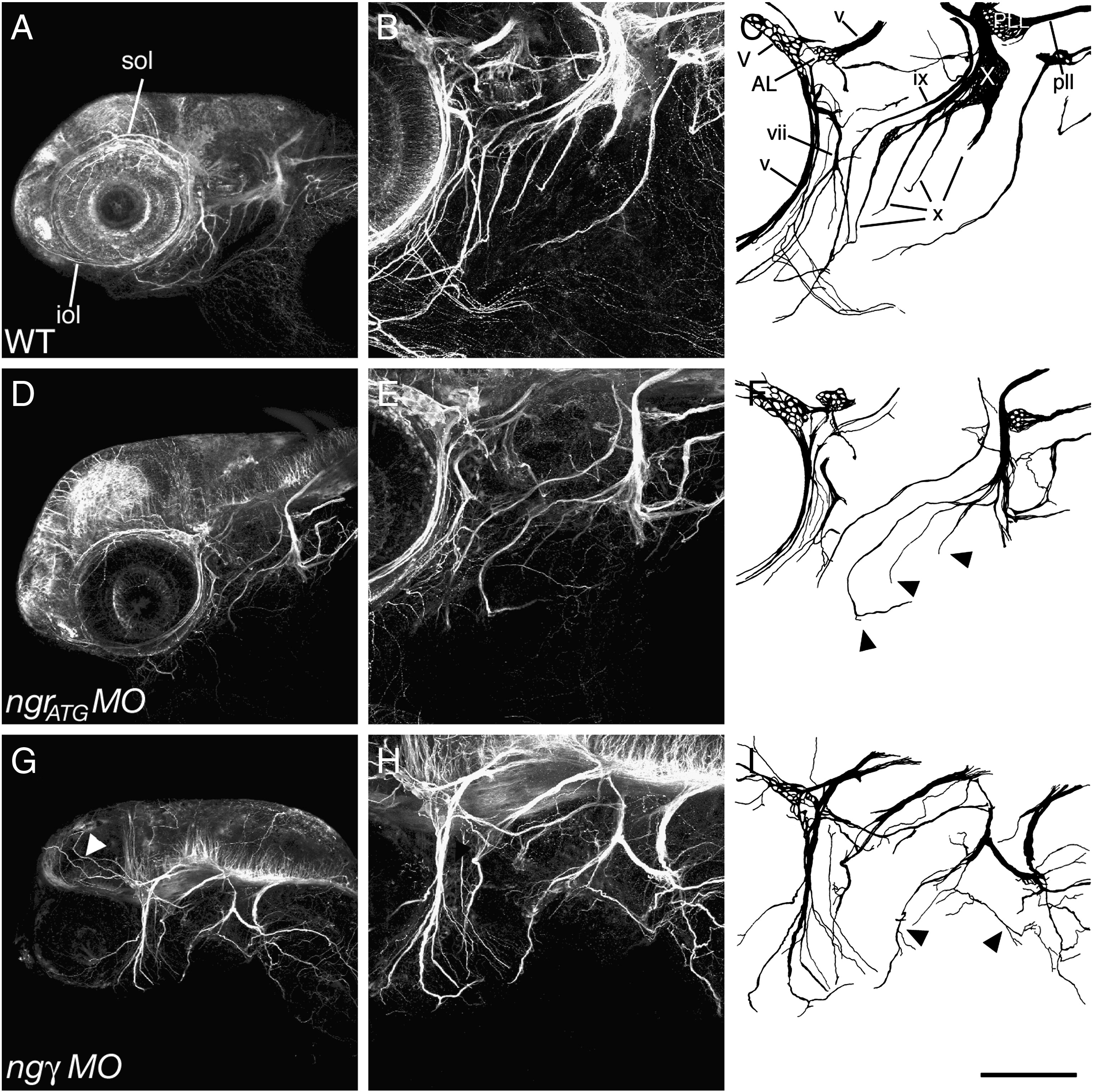
Fig. 3 Loss of Nogo-γ or Ngr function disrupts cranial nerve projections. (A–C) Anti-acetylated tubulin immunostaining at 3 dpf reveals highly stereotyped innervation of the larval head, allowing the identification of many cranial nerves (Higashijima et al., 2000). Injection of 5 ng of (D–F) ngrATG MO or (G–I) nogo-γ MO results in the disruption of the cranial innervation pattern. Many nerve branches were significantly thinner, shorter, or even absent. In addition, nerve trajectories were disturbed and axons projected into aberrant target areas (arrowheads). (A, B, D, E, G, H) Maximum intensity projections of side view confocal z-series of whole-mount zebrafish larvae. (B, D, F) are higher magnifications of the area caudal to the eye shown in (A, C, E), and depicted schematically in (C, F, I). iol = infraorbital lateral line nerve, sol = supraorbital lateral line nerve, V = trigeminal ganglion, v = trigeminal nerve, AL = anterior lateral line ganglion, vii = facial nerve, ix = hypoglossal nerve, X = vagal ganglion, x = branches of the vagal nerve, PLL = posterior lateral line ganglion, pll = posterior lateral line nerve. Scale bar A, C, E = 200 μm; B, D, F = 100 μm.
Reprinted from Molecular and cellular neurosciences, 40(4), Brösamle, C., and Halpern, M.E., Nogo-Nogo receptor signalling in PNS axon outgrowth and pathfinding, 401-409, Copyright (2009) with permission from Elsevier. Full text @ Mol. Cell Neurosci.