
Image
|
Figure Caption
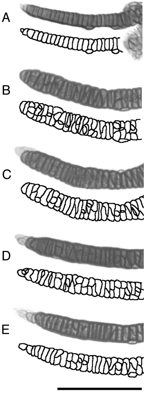
Fig. 6 Disruption of the SY cell stack during growth. Alcian green-labeled flat mounts, and accompanying tracings of the chondrocyte outlines. (A) An example at day 4. The SY cells are neatly stacked (compare with other examples in Figs. 1D, 2, and 3). (B, C) and (D, E): The left–right pairs of two larvae at day 8. Cellular addition variably disrupts the early stacks. The cells in many regions are substantially overlapped. Scale bar: 100 μm.
Acknowledgments
This image is the copyrighted work of the attributed author or publisher, and
ZFIN has permission only to display this image to its users.
Additional permissions should be obtained from the applicable author or publisher of the image.
Reprinted from Developmental Biology, 203, Kimmel, C.B., Miller, C.T., Kruse, G., Ullmann, B., BreMiller, R.A., Larison, K.D., and Snyder, H.C., The shaping of the pharnygeal cartilages during early development of the zebrafish, 245-263, Copyright (1998) with permission from Elsevier. Full text @ Dev. Biol.