
|
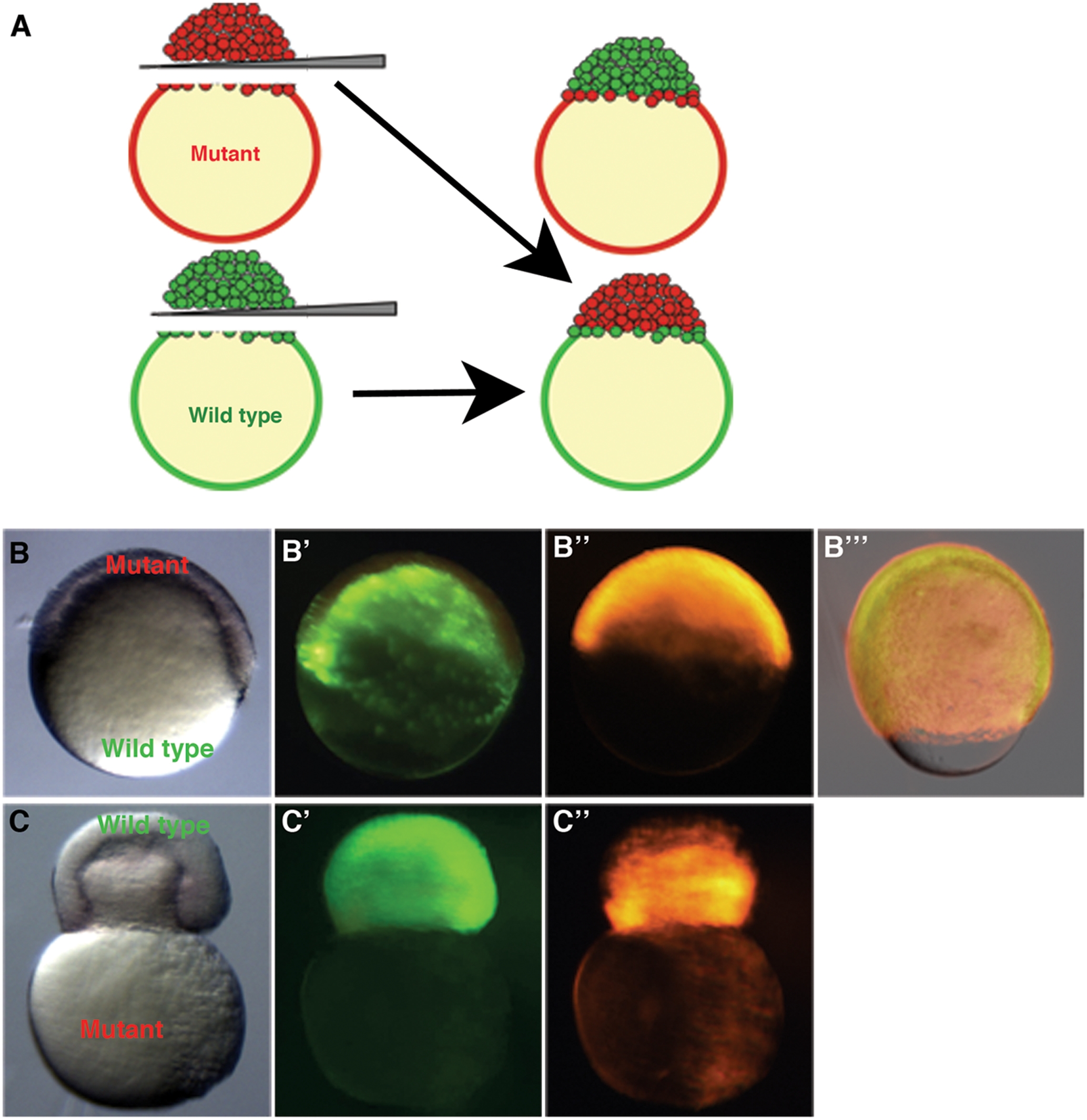
Fig. 5 Whole blastoderm transplants show yolk cell domain of function.
Embryos were injected with rhodamine-dextran (bbp) or sytox green dye (WT) at the 1-cell stage. Blastoderms were separated from yolks and re-adhered creating chimeric embryos. (A) Schematic representation of the blastoderm transplant. A chimeric embryo (B–B′″) containing a WT yolk (B′) and bbp blastoderm (B″) progresses through epiboly properly (B′″, 80% epiboly). A chimeric embryo (C–C″) containing a WT blastoderm (C′) and bbp yolk (C″) constricts at 50% epiboly, showing that Bbp functions in the yolk cell during epiboly. Bright field images of chimeric embryos, B, C. WT derived tissue, B′, C′. bbp derived tissue, B″, C″. Merge of images at 80% epiboly in B′″.