
Image
|
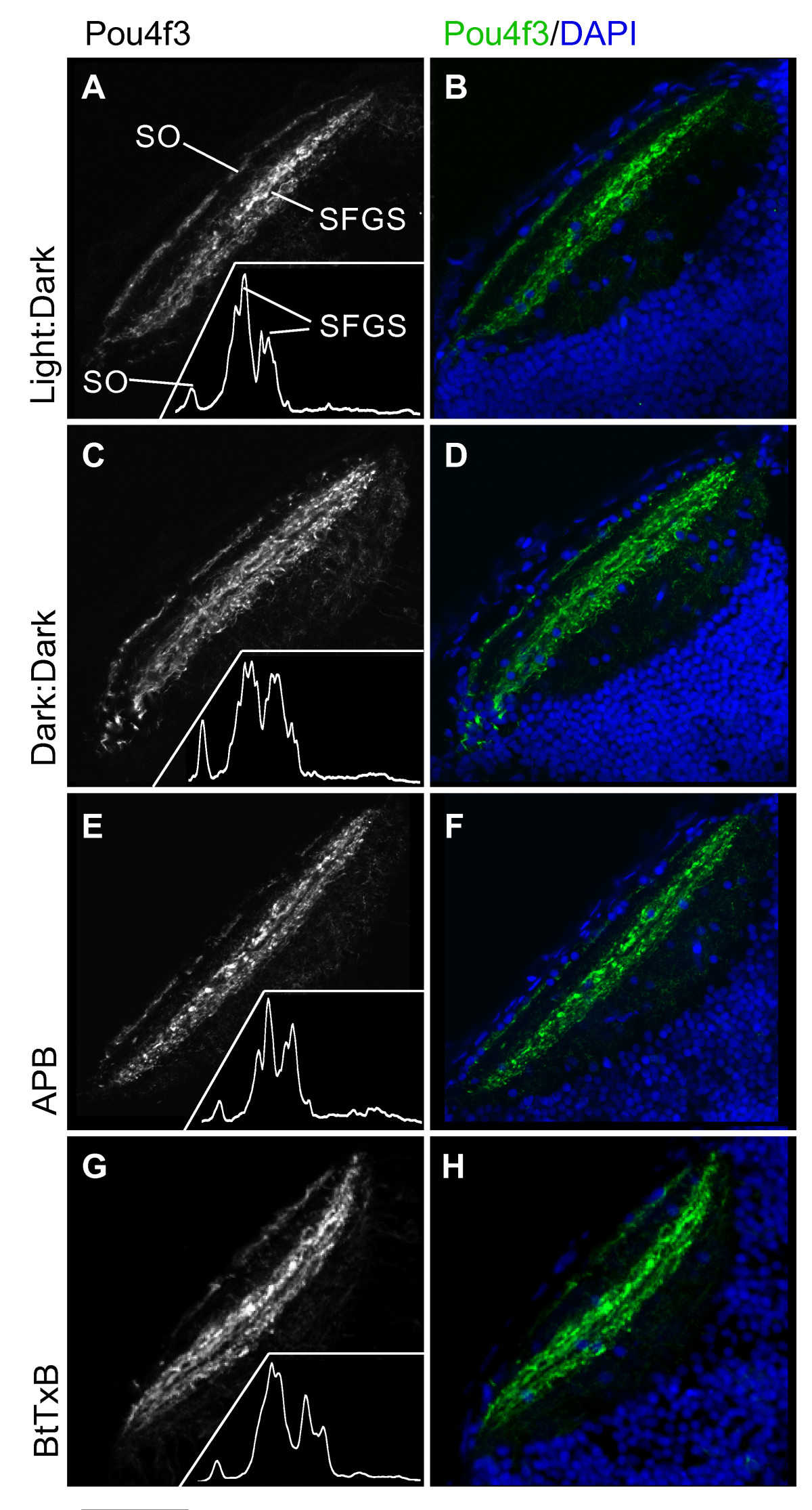
Figure Caption
Fig. 7 Dark-reared, APB-treated, and BtTxB-injected larvae show proper GC axon targeting to tectal laminae. Horizontal sections of 5 dpf larval tecta showing Pou4f3:mGFP+ GC axons innervating the optic tectum, imaged by confocal microscopy. (A, C, E, G) Pou4f3:mGFP+ axons innervate the SO and two sublaminae of the SFGS. Insets: densitometric traces across the tectal neuropil, from superficial to deeper layers. (B, D, F, H) Same images of Pou4f3+ axons (green), with DAPI labeling (blue) to show the cell body and neuropil regions of the tectum. Scale bar 50 μm.
Acknowledgments
This image is the copyrighted work of the attributed author or publisher, and
ZFIN has permission only to display this image to its users.
Additional permissions should be obtained from the applicable author or publisher of the image.
Full text @ Neural Dev.